
What this is
- This research investigates how traumatic brain injury (TBI) affects nucleolar dynamics in Drosophila, particularly in relation to aging.
- Nucleolar enlargement, associated with increased ribosome biogenesis, is a hallmark of aging and is influenced by the .
- The study shows that young flies experience significant nucleolar enlargement after TBI, while older flies do not, indicating age-dependent responses.
Essence
- TBI induces rapid nucleolar enlargement in young Drosophila, but older flies show minimal changes. Inhibition of the TOR pathway reduces early mortality after TBI, suggesting a link between nucleolar dynamics and injury outcomes.
Key takeaways
- Young Drosophila exhibit a 28.0% increase in nucleolar area after TBI, indicating heightened ribosome biogenesis. This enlargement persists for weeks, suggesting a prolonged cellular response to injury.
- Older flies show little to no nucleolar enlargement post-injury, with a 5.6% reduction in nucleolar area at 5 days after TBI. This highlights the diminished cellular response to injury with age.
- Pharmacological inhibition of the TOR pathway significantly reduces early mortality by 30.6% with rapamycin, indicating that nucleolar expansion contributes to adverse outcomes following TBI.
Caveats
- The study primarily uses a Drosophila model, which may limit the generalizability of findings to humans. Further research is needed to confirm these mechanisms in mammalian systems.
- While the study shows correlations between TBI, aging, and nucleolar size, causative mechanisms remain to be fully elucidated. Direct measurements of nucleolar size after TOR inhibition are necessary.
Definitions
- nucleolus: A nuclear structure where ribosomal RNA is processed and ribosomal subunits are assembled, reflecting cellular stress and ribosome biogenesis.
- TOR signaling pathway: A cellular pathway that regulates growth and metabolism, influencing nucleolar size and ribosome production, particularly during aging.
Simplified
Introduction
Traumatic brain injury (TBI) affects approximately 70 million people worldwide each year [1,2]. However, its underlying mechanisms remain poorly understood due to the complex, evolving cascade of secondary cellular and molecular responses influenced by both genetic and environmental factors [3 –5]. Emerging evidence suggests that secondary responses may impair recovery by accelerating normal aging processes [6,7]. Indeed, several hallmarks of aging, such as chronic inflammation, oxidative stress, mitochondrial dysfunction, and neurodegeneration, are exacerbated by TBI [6 –11].
Another hallmark of aging is altered nucleolar structure and function, which reflects changes in ribosome biogenesis and cellular stress responses. Nucleoli are specialized nuclear compartments where ribosomal DNA (rDNA) is transcribed, ribosomal RNA (rRNA) is processed, and ribosomal subunits are assembled [12,13]. They are composed of three distinct regions: the fibrillar center (FC), the dense fibrillar component (DFC), and the granular component. (GC) [14]. The DFC contains fibrillarin, a protein essential for rRNA processing. In many cell types, nucleolar size, as indicated by fibrillarin staining, increases with age [15,16]. For example, in human fibroblasts, nucleolar size correlates positively with donor age [17]. Furthermore, in C. elegans, knockdown of fibrillarin reduces nucleolar size and extends lifespan, supporting a causal link between nucleolar expansion and aging [16,18].
The target of rapamycin (TOR) signaling pathway plays a central role in regulating nucleolar size during aging [19,20]. Age-related increases in nutrient and growth factor signaling, combined with mitochondrial dysfunction, can contribute to aberrant TOR activation, which in turn promotes rDNA transcription and ribosome biogenesis. Pharmacological inhibition of TOR by rapamycin reduces nucleolar size and extends lifespan in multiple model organisms [16,19,20]. These findings raise the possibility that TBI may increase nucleolar size by accelerating age-related processes. In support of this idea, treatment with rapamycin improves various pathological outcomes of TBI in rodents [21 –28].
To investigate whether TBI affects nucleolar size in the brain, we used a Drosophila melanogaster model in which closed-head injury and associated polytrauma are induced with a High-Impact Trauma (HIT) device [29,30]. We previously showed that TBI outcomes in flies are strongly age-dependent and engage conserved aging pathways [29,31 –33]. Specifically, early mortality (death within 24 h of injury) increased with age at the time of injury and correlated with the median lifespan of different inbred fly lines [31,32]. Flies from longer-lived lines had lower early mortality than shorter-lived lines when injured at the same chronological age. Similarly, genetic and environmental interventions that extended lifespan also reduced early mortality after TBI. Additionally, we found that TBI in flies triggered innate immune system activation [32,34,35] and increased intestinal permeability [31,36], which are markers of impending death during normal aging [37], and caused severe neurodegeneration when older flies were injured [29]. These findings establish Drosophila as a powerful model for studying age-dependent TBI outcomes.
Using immunofluorescence microcopy, we found that TBI in young flies (3−6 days old) caused a substantial increase in brain nucleolar size at 1 day post-injury that was maintained over the following 40 days. In contrast, injury of older flies (26−29 or 47−50 days old) resulted in little or no nucleolar enlargement at 1 day after injury and smaller nucleoli than uninjured controls by 5 days post-injury. Furthermore, treatment of young flies with rapamycin or RapaLink-1, another TOR inhibitor, reduced early mortality after TBI. These findings support the idea that TBI accelerates aging-related increases in ribosome biogenesis, contributing to adverse outcomes.
Results and discussion
TBI rapidly increases nucleolar size
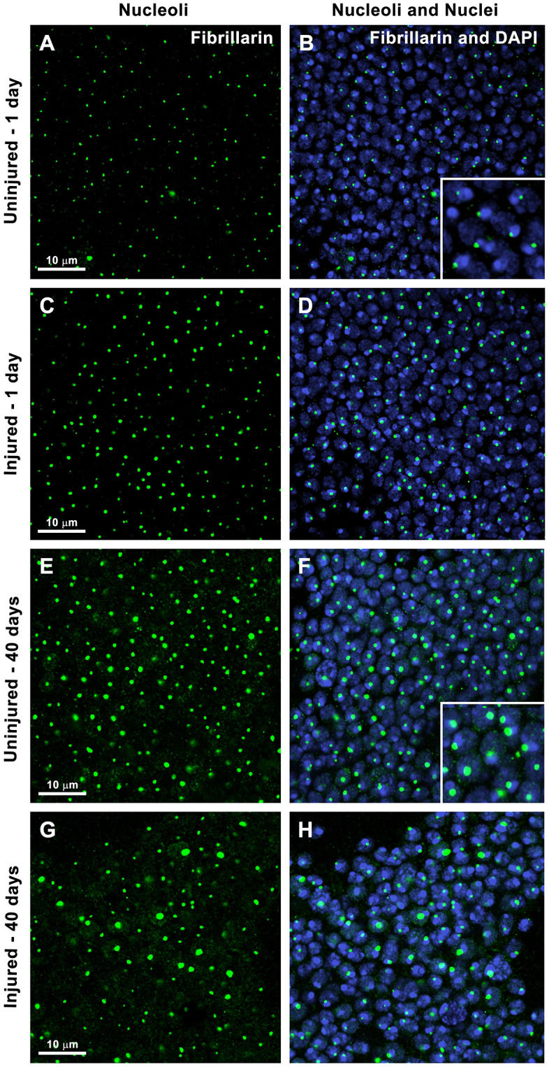
To investigate nucleolar dynamics following TBI, we used confocal immunofluorescence microscopy to assess nucleolar size in the central brain of injured and uninjured adult flies. These experiments used a standard laboratory fly strain, w1118. Mixed-sex flies (1:1, female:male) were injured at 3−6 days old using a HIT device; and age-matched, uninjured flies served as controls. Consistent with previous studies, injury by the HIT device caused 25% early mortality in this strain [29,31,32]. Brains from surviving flies and uninjured controls were stained with an antibody to fibrillarin to visualize nucleoli and DAPI (4',6-diamidino-2-phenylindole) to label nuclear DNA in neurons and glia (Fig 1).
In both injured and uninjured brains at 1 day post-injury, each nucleus contained a single nucleolus positioned adjacent to or partially embedded within a brightly DAPI-stained region (Figs 1B (inset) and 1D). Nucleoli in Drosophila form around multicopy rDNA arrays located in the heterochromatic regions of the X and Y chromosomes [38]. Because heterochromatin is rich in A-T base pairs, it binds DAPI with high affinity and appears strongly fluorescent [39]. In Drosophila, rDNA arrays on the sex chromosomes are typically paired, resulting in the formation of a single nucleolus per nucleus [38,40]. Thus, the data in Fig 1 are consistent with previous reports of nucleolar organization in Drosophila somatic cells.
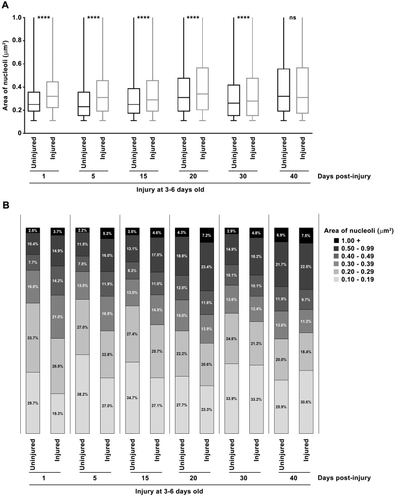
Visual inspection of images suggested that nucleoli were larger in injured flies compared with uninjured controls at 1 day after injury (compare Fig 1A and 1C). Quantitative analysis of at least 11 brains and 7,000 nuclei for each condition confirmed this observation: the median nucleolar area was significantly larger in injured flies (Fig 2A). Injury increased the median nucleolar area by 28.0%, from 0.25 μm2 in uninjured flies to 0.32 μm2 in injured flies. This shift was driven by a reduced proportion of small nucleoli (≤0.29 μm2) and an increased proportion of larger nucleoli (≥0.3 μm2) (Fig 2B). These findings suggest that TBI stimulates ribosome biogenesis, leading to nucleolar enlargement. However, it remains to be determined whether rRNA and ribosomal protein synthesis, and thus overall translational capacity, are elevated. Further work is also needed to identity which cell types exhibit enlarged nucleoli. Given that 17.2% of cells showed increased nucleolar size at 1 day post-injury (Fig 1B), whereas only 10% of brain cells are glia [41], the effect likely occurs in both neurons and glia or exclusively in neurons. To our knowledge, this is the first report showing that TBI alters nucleolar size, adding to the growing evidence TBI elicits cellular responses similar to those observed during normal aging [6,7,9 –11].

TBI and aging both increase nucleolar size in Drosophila brain. Representative confocal microscopy images of the central brain from uninjured (A, B, E, and F) and injured (C, D, G, and H) mixed-sex, 3-6 day old,flies at 1 day (A-D) and 40 days (E-H) post-injury. (B and F) Insets show magnified views, highlighting nucleoli located next to bright DAPI-stained spots. Nuclei are labeled with DAPI (blue) and nucleoli are labeled with fibrillarin antibody (green). w 1118

TBI and aging promote nucleolar expansion. Nucleolar area was measured by fibrillarin staining of dissected brains from 3-6 day old, mixed-sex,flies that were either uninjured or injured. For each condition and time point, at least 11 brains and 7,000 nucleoli were examined. (A) Nucleolar area is shown as box-and-whisker plots, with the median (horizonal line), interquartile range (box), and minimum and maximum values (whiskers). p-values reflect comparisons between injured and uninjured flies using one-way analysis of variance (ANOVA) with Tukey's multiple comparison test. ns = not significant, ****p < 0.0001. (B) Distribution of nucleolar areas, shown as the percent of nucleoli falling within each of five size ranges (see figure key). w 1118
TBI accelerates age-associated nucleolar enlargement
To investigate the long-term effect of TBI on nucleolar dynamics, we used immunofluorescence microscopy of fibrillarin to examine nucleoli at 5, 15, 20, 30, and 40 days post-injury. In uninjured control flies, nucleoli were visibly larger in older flies (43–46 day old, i.e., 3–6 day old flies aged 40 days, Fig 1E) compared to young flies (4–7 day old, i.e., 3–6 day old flies aged 1 day, Fig 1A). This visual observation was supported by quantitative analysis: in uninjured flies, the median nucleolar area increased by 28.0%, from 0.25 μm2 at 1 day to 0.32 μm2 at 40 days (Fig 2A). This increase was non-linear, with decreases between days 1–5 and 20–30. Thus, the nucleolar enlargement seen 1 day after injury is similar in magnitude to the age-related increase that occurs over 40 days in uninjured flies. These findings raise the possibility that injured cells undergo senescence, a state marked by nucleolar enlargement [42] and induced by age-associated stresses as well as by increased rRNA transcription or decreased rRNA processing [43,44].
As flies aged, the difference in nucleolar area between injured and uninjured flies diminished. At 1 day post-injury, the proportion of nucleoli with an area ≥0.3 μm2 was 17.2% higher in injured flies than in uninjured controls. This difference steadily declined to 15.4, 9.2, 6.0, 4.0, and −3.1% at 5, 15, 20, 30, and 40 days post-injury, respectively (Fig 2B). Since the median nucleolar area remained relatively stable in injured flies over time (Fig 2A), the narrowing gap between injured and uninjured flies is attributable to the age-dependent increase in nucleolar size in uninjured flies.
TBI and aging increase cell-to-cell variation in nucleolar size
In both injured and uninjured flies, the area of nucleoli ranged from 0.1 to >1.0 μm2 (Fig 2A), reflecting a combination of biological heterogeneity and imaging effects – nucleoli appear largest when sectioned through the center and smaller near the surface. One day post-injury, the interquartile range (IQR) of nucleolar size (25th to the 75th percentile) was narrower in uninjured flies (0.19–0.36 μm2) than in injured flies (0.22–0.45 μm2), suggesting that injury increases cell-to-cell variation in nucleolar size (see boxes in Fig 2A). Aging showed a similar effect: visual inspection (Fig 1A vs. 1E; 1C vs. 1G) and IQR analysis (Fig 2A) revealed that variation in nucleolar size widened with age. In uninjured flies, the IQR expanded from 0.19–0.36 μm2 at 1 day to 0.19–0.56 μm2 at 40 days; in injured flies, it widened from 0.22–0.45 μm2 at 1 day to 0.17–0.57 μm2 at 40 days. These findings suggest that both TBI and aging activate signaling pathways that drive nucleolar enlargement, but the extent of enlargement differs among brain cells, possibly due to cell-to-cell variation in gene expression [45,46].
Age at the time of TBI influences the nucleolar response to injury
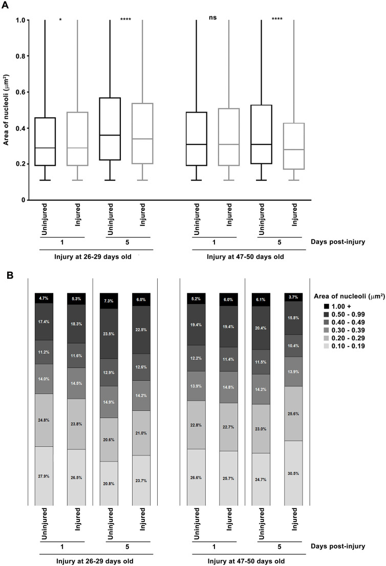
To investigate how aging influences the effect of TBI on nucleolar dynamics, we repeated the 1 and 5 day post-injury analyses using flies injured at 26–29 or 47–50 days old. Consistent with our prior studies [29,31 –33], early mortality increased with age at the time of injury: 30% for 26–29 day old flies and 45% for 47–50 day old flies. At 1 day post-injury, both 26–29 and 47–50 day old flies showed no difference in median nucleolar area between injured flies and uninjured controls (Fig 3A). This contrasts with the 28.0% increase observed in flies injured at 3–6 day old (Fig 2A). At 5 days post-injury, median nucleolar area was significantly reduced in both older groups – by 5.6% in 26–29 day old flies and by 9.7% in 47–50 day old flies (Fig 3A) – whereas nucleoli in injured 3–6 day old flies were 34.8% larger (Fig 2A). These reductions in older flies were associated with an increased proportion of nucleoli ≤0.29 μm2 and a corresponding decrease in those ≥0.3 μm2 (Fig 3B). These findings suggest that aging activates the molecular pathways that regulate nucleolar size to their maximum capacity, leaving no additional effect for TBI to exert.
TBI-induced changes in nucleolar dynamics may contribute to downstream age-related pathologies, including mitochondrial dysfunction, DNA damage, cellular senescence, and neurodegeneration. Ribosome biogenesis, a highly energy- and resource-demanding process, can strain mitochondrial capacity, impairing function [47]. In addition, expansion of the nucleolus beyond a critical size can compromise nucleolar integrity, leading to DNA damage through influx of the homologous recombination repair protein Rad52 and aberrant recombination [22]. Excessive rDNA transcription without coordinated ribosome biogenesis can drive cells into senescence [44]. Finally, reduced nucleolar size, as occurred days after injury of older flies (Fig 3), is seen in neurodegenerative diseases like Alzheimer's and Parkinson's [13,48 –51].

TBI has minimal acute effects on nucleolar expansion in older flies. Nucleolar area was determined by fibrillarin staining of dissected brains from mixed-sex,flies that were either uninjured or injured at 26-29 or 47-50 days old and aged 1 or 5 days. For each condition and time point, at least 11 brains and 7,000 nuclei were analyzed. (A) Nucleolar area is shown as box-and-whiskers plots, displaying the median (horizonal line), interquartile range (box), and minimum and maximum values (whiskers). p-values reflect comparisons between injured and uninjured flies using one-way ANOVA with Tukey's multiple comparison test. ns = not significant, *p < 0.05, ****p < 0.0001. (B) Distribution of nucleolar areas, shown as the percent of nucleoli falling within each of five size ranges (see figure key). w 1118
Inhibition of the TOR pathway reduces early mortality following TBI
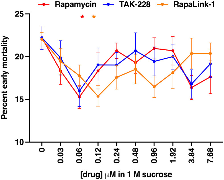
To test whether nucleolar expansion contributes to TBI outcomes, we assessed the effect of TOR pathway inhibition on early mortality. Mixed-sex, w1118 flies (0–7 days old) were injured and then fed the TOR inhibitors rapamycin, TAK-228, or RapaLink-1 delivered in 1 M sucrose across a 256-fold concentration range (0.03–7.68 μM). We have previously shown that feeding flies 1 M sucrose results in the same percent early mortality following TBI as flies fed standard fly food [31]. TAK-228, also known as MLN0128 and INK128, is an ATP-competitive TOR kinase inhibitor [52,53], and RapaLink-1 is a bifunctional molecule that chemically links rapamycin to TAK-228 [54]. Rapamycin and RapaLink-1 significantly reduced early mortality at 0.06 μM and 0.12 μM, respectively, with reductions of 30.6% and 30.2% (Fig 4). TAK-228 also showed a near-significant effect at 0.06 μM (p = 0.079). These findings suggest that TOR-dependent nucleolar expansion contributes to TBI pathogenesis. Because rapamycin and RapaLink-1 primarily inhibit mechanistic target of rapamycin complex 1 (mTORC1) rather than mTORC2 in flies, our findings further suggest that early mortality following TBI is mediated by mTORC1 [55]. However, although reduced early mortality following TOR inhibition is consistent with TOR-dependent nucleolar expansion, definitive proof will require directly measuring nucleolar size in flies treated with TOR inhibitors. Such measurements are necessary to establish a causal link between nucleolar enlargement and early mortality.
Mechanisms linking nucleolar expansion to TBI pathogenesis remain unclear. One possibility is that increased ribosome biogenesis elevates protein synthesis beyond the capacity of protein folding and quality control systems, resulting in protein aggregation and endoplasmic reticulum stress [56] – both observed after TBI [57,58]. Another is that the high energy demand of ribosome production [59] depletes energy in neurons already impaired by mitochondrial dysfunction [60,61], reducing their ability to maintain ion gradients and detoxify reactive oxygen species. Alternatively, nucleolar expansion may arise from an imbalance between ribosomal proteins and rRNA, leading to accumulation of unprocessed rRNA, ribosomal precursors, or orphan ribosomal proteins [62,63]. This can trigger nucleolar stress and cellular toxicity, effects that can be mitigated by TOR inhibition [64].
In summary, we found that TBI induced rapid nucleolar enlargement that persisted for weeks in young flies but was absent in older ones, indicating an age-dependent response (Figs 1–3). Both TBI and aging also increased cell-to-cell variation in nucleolar size (Figs 1 and 2). Notably, inhibition of TOR signaling – a key regulator of nucleolar size – reduced early mortality after TBI (Fig 4). These findings suggest that nucleolar enlargement contributes to TBI-related adverse outcomes and highlight Drosophila as a powerful model for dissecting the mechanisms that link nucleolar dynamics to TBI pathophysiology.

TOR inhibitors reduce early mortality after TBI in a dose-dependent manner. Mixed-sex,flies (0-7 days old) were fed rapamycin, TAK-228, or RapaLink-1 in 1 M sucrose following injury. Early mortality was measured as the percent of flies that died within 24 h post-injury. Data represent the mean ± standard error of the mean (SEM) from 12 replicates (60 flies each) for rapamycin, 12 replicates for TAK-228, and 9 replicates for RapaLink-1. Statistical comparisons of each drug treatment were performed using one-way ANOVA with Dunnett's multiple comparison test; *p < 0.05. w 1118
Materials and methods
Fly culturing and TBI
Flies were maintained at 25°C on a standard fly food containing cornmeal, molasses, and yeast [65]. The w1118 strain has been maintained in our lab for many years. Flies were collected at 0–3 days post-eclosion, aged as indicated, and subjected to TBI in groups of 60 using a HIT device (4 strikes, spaced 5 min apart), following the protocol described in Katzenberger et al., 2013 [29]. Control (uninjured) flies were handled identically but were not subjected to strikes. After treatment, both injured and uninjured flies were transferred to fresh food vials and maintained at 25°C until analysis. Early mortality was calculated as the percent of flies that died within 24 h post-injury, adjusted by subtracting the percent mortality in the uninjured group. Flies were considered dead if they remained immobile following gentle agitation.
Confocal immunofluorescence microscopy and quantitation of nucleolar area
Brains were dissected under a light microscope in a drop of fresh, ice cold 4% formaldehyde on a Sylgard plate (Dow Inc.) with #5 Dumont forceps (Fine Science Tools) and transferred to an Eppendorf tube containing 1 ml 4% formaldehyde on ice for approximately 30 min. Brains were washed 3 x 20 min in 1.5 ml 1X phosphate buffered saline (PBS) with 0.3% Triton-X (PBS-T), blocked in 500 μl PBS-T with 5% normal goat serum (Sigma) overnight at 4°C. Block was removed and 1.5 ml fibrillarin antibody (1:500 in PBS-T, 1:100, Cytoskeleton (AFB01)) was added and incubated overnight at 4°C. Brains were washed 3 x 20 min in 1.5 ml PBS-T at room temperature. Fluorescently labeled secondary antibody (α-mouse Alexa Fluor 488, 1:500 in PBS-T, Invitrogen) was added and incubated overnight at 4°C. Brains were washed 3 x in 1.5 ml PBS-T at room temperature. The first wash (10 min) contained DAPI (1 mg/mL in H2O, diluted 1:1000 in PBS-T, Santa Cruz Biotechnology), and the second and third washes (10 min each) were only PBS-T. During washes and incubations, tubes were placed in a light tight box and agitated. Brains were mounted in Vectashield (Vector Laboratories) and imaged at 100x magnification on a Nikon A1R-SI+ confocal microscope (Optical Imaging Core, University of Wisconsin, Madison, WI).
Nikon Elements Imaging Software (Optical Imaging Core, University of Wisconsin, Madison, WI) was used to determine the area of individual nucleoli in the central brain. 100x single channel confocal images were used for this analysis of at least 11 brains and 7,000 nucleoli. In the 488 channel, fibrillarin-stained nucleoli were selected through thresholding to create a binary mask. The software calculated the area based on the number of pixels within each focus in the binary mask, converting pixels to calibrated units (μm2). Approximately half of the brains were imaged from the posterior side and the other half from the anterior side. Nucleoli were sorted by area using Microsoft Excel, and those >0.1 μm2 were counted. GraphPad Prism (version 10.5.0) was used for graphing and statistical analyses of the data in Figs 2 and 3.
Drug treatment
Injured and uninjured, mixed-sex, 0–7 day old, w1118 flies were transferred to vials containing a filter paper disc at the bottom absorbed with 200 μl of 1 M sucrose or rapamycin (Sigma), TAK-228, or RapaLink-1 (TAK-228 and RapaLink-1 were supplied by Douglas Wassarman and Kevan Shokat, University of California-San Francisco) at the indicated concentrations in 1 M sucrose (Fig 4). For each concentration and condition, 12, 12, and 9 independent samples of 60 flies were examined for rapamycin, TAK-228, and RapaLink-1, respectively. GraphPad Prism (version 10.5.0) was used for graphing and statistical analyses of the data in Fig 4.