
What this is
- This research investigates the role of astrocyte senescence in the aging hippocampus, specifically its impact on synapse formation.
- Using the SAMP8 mouse model, the study identifies early signs of senescence in astrocytes and their reduced ability to promote synaptogenesis.
- The findings focus on the loss of (TSP-1), a key protein for synapse formation, in senescent astrocytes.
Essence
- Astrocyte senescence leads to impaired synapse formation in the aging hippocampus due to reduced levels. Restoring TSP-1 can rescue synaptogenic function.
Key takeaways
- Senescent astrocytes in the SAMP8 mouse model exhibit increased SA-β-gal activity, indicating early signs of aging. This accumulation correlates with reduced synaptic function.
- levels are significantly lower in senescent astrocytes, impairing their ability to support synaptogenesis. Supplementing with TSP-1 can restore this function.
- The study suggests that targeting astrocyte senescence and TSP-1 levels may offer therapeutic strategies to mitigate synaptic loss and cognitive decline in aging.
Caveats
- The study relies on a specific mouse model (SAMP8), which may not fully represent human aging processes. Further research is needed to validate findings in human contexts.
- The focus on TSP-1 as a singular factor in synaptogenesis may overlook other potential contributors to synaptic dysfunction in aging.
Definitions
- Cellular senescence: An irreversible state of cell cycle arrest that negatively impacts tissue function, often associated with aging.
- Thrombospondin-1 (TSP-1): A protein secreted by astrocytes that plays a critical role in synapse formation and neuronal development.
Simplified
Introduction
Aging is a complex process that involves the progressive deterioration of the organism. It is characterized by the accumulation of damage throughout life and the gradual loss of tissue and organ function (Kirkwood 2017). Scientific discoveries in recent decades have identified a set of interconnected processes, known as the "hallmarks of aging," considered as a starting point for exploring the foundations of aging and for developing treatments to counteract age‐related functional decline (López‐Otín et al. 2023).
Cellular senescence, a hallmark of aging, stands out among the causal processes of organismal aging (Campisi 2005). In proliferating cells, senescence has been defined as a state of irreversible cell cycle arrest that involves genetic, morphological, and functional alterations with a marked negative impact on tissue function (Cohen and Torres 2019; Dos Santos et al. 2024). Both proliferating and non‐proliferating cells can undergo senescence in response to a variety of internal and external factors, including cytotoxic stimuli, oxidative stress, proteasome inhibition, DNA damage, and alterations in cellular communication systems, due to paracrine signals released by neighboring senescent cells (Takai et al. 2003; Torres et al. 2006; Pricola et al. 2009; Bitto et al. 2010; Rodier et al. 2011; Acosta et al. 2013; Clevers and Watt 2018). Cellular senescence can be detected through a variety of senescence markers such as increased expression of the cyclin‐dependent kinase (CDK) inhibitors CDKN2A/p16INK4a and CDKN1A/p21CIP1 (Evans et al. 2003; Bhat et al. 2012; Yu et al. 2017; Idda et al. 2020), changes in nuclear structure and decreased levels of the nuclear lamina protein Lamin B1 (Freund et al. 2012; Matias et al. 2021), increased lysosomal mass linked to the detection of elevated levels of senescence‐associated β‐galactosidase activity (SA‐β‐gal) at pH 6 (Dimri et al. 1995; Evans et al. 2003; Piechota et al. 2016; Yu et al. 2017), and increased secretion of proinflammatory chemokines, cytokines, proteases and growth factors, collectively known as the senescence‐associated secretory phenotype (SASP) (Coppé et al. 2008; Bhat et al. 2012; Cohen et al. 2017; Gorgoulis et al. 2019). Despite the close association between cellular senescence and age‐related tissue dysfunction, the role of cellular senescence in brain aging remains poorly explored.
The SAMP8 (Senescence‐Accelerated Mouse Prone 8) mouse is a non‐transgenic model of accelerated aging widely used to study brain aging. Compared to the control SAMR1 (Senescence‐Accelerated Mouse Resistant 1) strain, SAMP8 animals show early signs of motor impairment, muscle weakness, and memory deficits, as observed in human aging (Dacomo et al. 2024). At the brain level, SAMP8 animals show elevated oxidative stress, chronic inflammation, altered microglia, decreased neurogenesis, and synaptic and neuronal dysfunction, among other symptoms (Tanisawa et al. 2013; Griñan‐Ferré et al. 2016; Yanai and Endo 2016; Díaz‐Moreno et al. 2018; Fernández et al. 2021). Furthermore, the SAMP8 model develops neurodegenerative changes with age and has been proposed as a relevant model for the study of age‐related diseases such as late‐onset Alzheimer's disease (Morley 2002; Pallas et al. 2008; Akiguchi et al. 2017). Therefore, SAMP8 stands as a highly useful tool for studying the mechanisms of brain aging and exploring potential therapeutic interventions.
Astrocytes are the most abundant cell type in the brain (Miller 2018). They fulfill fundamental functions in brain homeostasis, such as blood–brain barrier (BBB) maintenance, immune signaling, ionic level control, neurotrophin secretion, synapse formation, and neurotransmitter recycling (Chen et al. 2006; Phatnani and Maniatis 2015; Cheng et al. 2016; Miller 2018). Astrocyte senescence has been proposed to contribute to the age‐related loss of astrocyte function (Cohen and Torres 2019; Gorgoulis et al. 2019). However, research aimed at uncovering the impact of astrocyte senescence on the synaptic loss associated with aging and neurodegenerative diseases is very scarce. Here, we have taken advantage of the SAMP8 model to specifically interrogate the contribution of astrocyte senescence to synapse loss in the aged hippocampus, a paradigmatic region for synaptic plasticity studies in the context of learning and memory. Taking advantage of the SA‐β‐galactosidase staining, we show that senescence increases in astrocyte‐enriched layers of the SAMP8 hippocampi and in acutely isolated SAMP8 hippocampal astrocytes. We also employ two methodologies for exploring the synaptogenic capacity of senescent astrocytes: (i) isolation of adult hippocampal astrocytes from SAMP8 and SAMR1 based on the astroglial cell surface marker ACSA‐2, followed by primary culture in defined medium, and (ii) differentiation of hippocampal neural stem cells (NSCs) derived from adult SAMP8 and SAMR1 mice into astrocytes. We characterize the astrocytes using a variety of senescence markers and assess their functionality in synaptogenesis assays, employing primary hippocampal neuronal cultures. Our results show that hippocampal SAMP8 astrocytes progressively accumulate senescence marks, downregulate the production of the pro‐synaptogenic signal thrombospondin‐1 (TSP‐1), and precociously lose their synaptogenic activity during aging. The decrease in TSP‐1 levels parallels the excitatory synapse reduction observed in all astrocyte‐enriched layers of the SAMP8 hippocampus.
Experimental Procedures
Animals
The accelerated senescence SAMP8/TaHsd model and the senescence‐resistant control SAMR1/TaHsd strain (Takeda et al. 1981) were used in this study. Mice were purchased from Inotiv Inc. and housed in the animal facility of the Biomedicine Institute of Valencia. Experiments were performed in male young (2 months, 2‐m), middle‐aged (6 months, 6‐m), and old (10 months, 10‐m) animals. For hippocampal neuron culture, RjOrl:SWISS wild‐type mice were purchased from Janvier Labs, and timed‐pregnant mice (E18.5) were bred in the animal facility of the Biomedicine Institute of Valencia. Mice were sacrificed by cervical dislocation for primary cultures, and by anesthetic overdose prior to perfusion for immunohistochemistry techniques. Animals were bred under controlled temperature conditions, 12 h light/dark cycle and water and food ad libitum. All experimental procedures and handling were in accordance with the European Union Council guidelines (2010/63/EU) and the Spanish regulation (RD53/2013). Procedures were approved by the Ethics and Animal Welfare Committee (CEEA) of the Biomedicine Institute of Valencia and CSIC (CEEA references: 2023‐VSC‐PEA‐0212 and 2024‐VSC‐PEA‐0094).
Primary Culture of‐2andAstrocytes ACSA SAMR1 SAMP8 +
2, 6, and 10‐m SAMR1 and SAMP8 mice were euthanized by cervical dislocation and hippocampi were dissected from the brain. Hippocampal cell suspension was obtained using the Adult Brain Dissociation Kit (Miltenyi, 130‐107‐677) in combination with the gentleMACS Octo Dissociator (Miltenyi, 130–095‐937). ACSA‐2+ cells (astrocytes) were isolated by magnetic cell separation (MACS) method following the manufacturer's procedure and protocols (Miltenyi Biotec, 130‐097‐679). ACSA‐2+ cells were then seeded (200,000 cells) in 24‐well poly‐D‐lysine (Sigma‐Aldrich, 27964‐99‐4) and laminin‐coated plate (Sigma‐Aldrich, L2020), as recommended by Miltenyi Biotec for primary astrocyte cultures from the adult brain. Astrocytes were maintained in the defined commercial medium AstroMACS (Miltenyi Biotec, 130‐117‐031) supplemented with 2 mM L‐glutamine (Lonza, 17‐605C) and 100 μ/mL penicillin–streptomycin (Lonza, 17‐603E). Astrocytes were maintained for 14 days in vitro (DIV) and every other day, half of the medium was replaced. On the final day, cells were fixed with 2% paraformaldehyde (PFA) (PanReac, 141451). The cultures were maintained in an incubator at 37°C in controlled humidity at 5% CO2.
Primary Culture of Hippocampal Neurons From Wild‐Type Mice
For the primary culture of hippocampal neurons, 18.5‐day old embryos (E 18.5) from wild‐type mice were used. Embryos were decapitated and hippocampal cell suspension was obtained using the Neural Tissue Dissociation Kit (Miltenyi, 130‐092‐628) in combination with the gentleMACS Octo Dissociator (Miltenyi, 130‐095‐937). Neurons were isolated by MACS using the Neuron Isolation Kit (Miltenyi Biotec, 130‐115‐390). Neurons were seeded (150,000 cells) onto poly‐D‐lysine and laminin‐coated 24‐well plate and maintained for 11 DIV. Neurobasal medium (Thermo scientific, 21103049) supplemented with B‐27 (1×) (Thermo scientific, 17504044), 2 mM L‐glutamine and 100 μ/mL penicillin–streptomycin was used. On the first day post‐plating, AraC (2 μM) was added to reduce non‐neuronal contamination. The cultures were maintained in an incubator at 37°C in controlled humidity at 5% CO2.
At 11 DIV, neuron medium was totally replaced by ACM from Ast‐Diff or half replaced by ACSA‐2+ ACM during 3 h. Gabapentin (GBP, MedChemExpress, HY‐A0057) and TSP‐1 (MedChemExpress, HY‐P701325) dosages were based on Cheng et al. (2016) procedures. GBP (32 μM) was added to neurons 30 min before ACM treatment and maintained 3.5 h with the neurons. TSP‐1 (250 ng/mL) was added directly diluted with the ACM during 3 h. Finally, neurons were fixed with 2% paraformaldehyde (PFA) and synapses were quantified.
Culture of Differentiated Astrocytes (Diff‐Astrocytes) Derived FromandNeural Stem Cells () SAMR1 SAMP8 NSCs
SAMR1 and SAMP8 NSCs were isolated from the hippocampal dentate gyrus of 2‐m mice and expanded in the presence of mitogens following Babu's protocol (Babu et al. 2011). These cells were maintained in adherent culture with Neurobasal medium supplemented with B‐27 (1×), L‐glutamine (2 mM), penicillin–streptomycin (100 μ/mL), fibroblast growth factor 2 (FGF2 20 ng/mL) (PeproTech, 100‐18B) and epidermal growth factor (EGF 20 ng/mL) (PeproTech, AF‐315‐09). For cellular assays, experiments were conducted using comparable passage (P) number for both models, and cells were discarded at approximately P26. To obtain SAMR1 and SAMP8 Diff‐Astrocytes, NSCs were seeded (150,000 cells) in differentiation medium, consisting of Neurobasal medium supplemented with B‐27 (1×), L‐glutamine (2 mM), penicillin–streptomycin (100 μ/mL) and Fetal Bovine Serum (FBS at 5%) (Thermo scientific, 10100147) for a period of 8–11 days. On Day 4, half of the medium was replaced with fresh differentiation medium. Cultures were maintained in an incubator at 37°C in controlled humidity at 5% CO2. For immunofluorescence staining, 8 DIV cells were fixed on coverslips in 2% PFA for 10 min.
SAMP8 Diff‐Astrocytes were co‐transfected at day 11 with pcDNA3 mTSP1 and pMaxGFP, or with empty pcDNA3 and pMaxGFP as a control, using Lipofectamine 2000 (Fisher scientific, 15338‐030). pcDNA3 mTSP1 was a gift from Paul Bornstein (Addgene plasmid #12405; http://n2t.net/addgene:12405↗; RRID: Addgene_12405). 24 h after the transfection, the medium was changed to remove Lipofectamine. Cells were processed for RNA extraction or astrocyte conditioned medium collection 2 days later.
Astrocyte Conditioned Medium () Collection and Treatment ACM
Diff‐Astrocyte cultures were seeded in 24‐well plates and maintained 11 DIV. The culture medium was then completely removed and replaced with fresh differentiation medium for a 24‐h conditioning. After this time, the conditioned medium was collected and centrifuged at 300 g for 5 min to remove cellular traces. ACM from Diff‐astrocytes and transfected Diff‐astrocytes was immediately applied for 3 h to the hippocampal neurons (via full media change), or stored at −20°C.
On the other hand, ACM collection from primary ACSA‐2+ astrocyte cultures required a different procedure. Astrocytes were seeded in 24‐well plates and half of the AstroMACS medium was collected and replaced every 48 h for a period of 14 days. The medium was collected from each well, pooled, and centrifuged at 300 g for 5 min to remove cellular traces. For neuronal treatment, half of the neuronal culture medium was replaced during 3 h with ACSA‐2+ SAMR1 or SAMP8 ACM.
Senescence‐Associated β‐Galactosidase (‐β‐Gal) Reaction SA
The SA‐β‐gal reaction was performed following the protocol established by Debacq‐Chainiaux et al. (2009) with minor modifications. This reaction was performed in the same way for hippocampal tissues and for cell culture models. For cell cultures, the cells were fixed with 2% formaldehyde and 0.2% glutaraldehyde, followed by 3 washes with 0.1 M phosphate buffer (PB). The β‐galactosidase staining solution was then prepared and the samples were incubated with it for 5 h at 37°C with humidity. Primary antibodies were then applied to label astrocytes and nuclei with DAPI (Sigma‐Aldrich, D9542). Finally, the number of SA‐β‐gal positive cells was quantified as a percentage of the total cells or astroglial cells photographed per field. For tissue, the sections were completely immersed in the staining solution for 5 h and subsequently photographed. SA‐β‐gal signal intensity was normalized to the quantification area (μm2).
Immunocytochemistry of Astrocytes and Neurons
To perform immunofluorescence techniques, cells were pre‐fixed with 2% PFA for 10 min, followed by washing with 0.1 M PB. Subsequently, the samples were incubated at room temperature for 1 h with a blocking buffer composed of 0.1 M PB, 10% FBS, and 0.5% Triton X‐100. The primary antibody was then diluted in this blocking solution. This incubation was performed at 4°C for 24 h. The following day, cells were incubated with the secondary antibody, diluted in 0.1 M PB, for 2 h. DAPI was used to label nuclei. Primary antibodies: mouse anti‐GLAST (1:50; Miltenyi Biotec, 130‐119‐161), rabbit anti‐ATP1B2 (1:200; Alomone Labs, ANP‐012), guinea pig anti‐MAP2 (1:1000; Synaptic Systems, 188,004), rabbit anti‐VGLUT1 (1:300; GeneTex, GTX133148), mouse anti‐PSD95 (1:500; Thermo scientific, MA1045), rabbit anti‐GFAP (1:500; Sigma‐Aldrich, G3893), mouse anti‐TSP‐1 (1:50; Santa Cruz, sc‐59887) and guinea pig anti‐S100β (1:500; Synaptic Systems, 287004). Secondary antibodies: Alexa Fluor 488 anti‐mouse (1:500; Invitrogen, A21202), Alexa Fluor 555 anti‐mouse (1:500; Invitrogen, A31570), Alexa Fluor 488 anti‐rabbit (1:500; Invitrogen, A21206), Alexa Fluor 647 anti‐rabbit (1:500; Invitrogen, A31573), Alexa Fluor 488 anti‐guinea pig (1:500; Jackson, 706‐545‐148) and Alexa Fluor 647 anti‐guinea pig (1:500; Jackson, 706‐605‐148).
Immunohistochemistry of Mouse Hippocampal Tissue
The brains were processed with a Leica VT 1200 vibratome. Serial 40 μm sections comprising the entire hippocampus were collected. Before immunostaining, the tissue was washed three times for 5 min with 0.1 M PB to remove azide from the storage medium. The tissue was then incubated at room temperature for 1 h with a blocking buffer composed of 0.1 M PB, 10% FBS, and 0.5% Triton X‐100. The primary antibody was then diluted in the blocking buffer, and the sections were completely immersed in the mixture and kept shaking at 4°C for 24 or 48 h. The next day, the slices were then incubated for 2 h with the secondary antibody diluted in 0.1 M PB, protecting them from light. DAPI was used to label nuclei. Primary antibodies: rabbit anti‐VGLUT1 (1:300; GeneTex, GTX133148), mouse anti‐PSD95 (1:500; Thermo scientific, MA1045), rabbit anti‐GFAP (1:500; Sigma‐Aldrich, G3893), mouse anti‐TSP‐1 (1:50; Santa Cruz, sc‐59887), and guinea pig anti‐S100β (1:500; Synaptic Systems, 287004). Secondary antibodies: Alexa Fluor 555 anti‐mouse (1:500; Invitrogen, A31570), Alexa Fluor 488 anti‐rabbit (1:500; Invitrogen, A21206), Alexa Fluor 647 anti‐rabbit (1:500; Invitrogen, A31573) and Alexa Fluor 488 anti‐guinea pig (1:500; Jackson, 706‐545‐148).
Confocal Imaging and Image Analysis
Cells and tissue were observed with a Leica TCS SP8 confocal microscope and LSM 980 confocal microscope with ZEISS Airyscan detector. For the SA‐β‐gal reaction in cells, the images were obtained on a Leica DM6 B Automatic Upright Microscope (Thunder) and for tissue a Leica Aperio Versa Digital Scanner was used. Tissue images were captured at 1024 × 1024 resolution with a 40× focal lens, tracing vertical maps to obtain representative areas of all hippocampal regions. Cell photographs were taken with a 40× or 63× focal lens at 512 × 512 resolution, with at least 5 representative photographs of each coverslip. The colocalization synapse analysis was performed by neuronal immunostaining of pre‐ and post‐synaptic markers (VGLUT1 and PSD95 respectively) and normalized to the total number of neurons (MAP2+) of the image, using the Puncta Analyzer plugin and protocol of analysis (Ippolito and Eroglu 2010), with Fiji ImageJ software.
Quantitative‐ RT qPCR
RNA was extracted from cell cultures using a column extraction kit (Cytiva, 25050071) and subsequently quantified using a NanoDrop spectrophotometer. cDNA was obtained using the PrimeScript RT Reagent Kit (Takara, RR037A), following the manufacturer's instructions. The qPCR reaction was performed using the TB Green Premix Ex Taq Kit (Takara, RR82LR) and a QuantStudio 5 thermal cycler (Applied Biosystems). The specific forward and reverse oligonucleotides were as follows:(5′–3′): Lmnb1: (F) CAACTGACCTCATCTGGAAGAAC, (R) TGAAGACTGTGCTTCTCTGAGC; Il1β: (F) CAGGCAGGCAGTATCACTCA, (R) TAATGGGAACGTCACACACC; Il6: (F) CAAAGCCAGAGTCCTTCAGAG, (R) TGGTCCTTAGCCACTCCTTC; Cdkn1a: (F) GGCAGACCAGCCTGACAGAT, (R) TTCAGGGTTTTCTCTTGCAGAAG; Cacna2d1: (F) CCAAATCTTCAGCCAAAGGAGC, (R) ATTGACAGGCGTCCATGTGT. mRNA expression levels were calculated using the 2‐ΔΔCT method, according to Livak and Schmittgen (2001). Sdha was used as a housekeeping gene for normalization: (F) AGAGGACAACTGGAGATGGCATT, (R) AACTTGAGGCTCTGTCCACCAA.
Glutamate Uptake Assay
Free glutamate in the culture medium from NSCs and SAMR1 or SAMP8 Diff‐Astrocytes was quantified using the commercial Glutamate Assay kit, following the manufacturer's instructions (Sigma‐Aldrich, MAK004). Glutamate levels were normalized with an MTT (3‐(4, 5‐dimethylthiazolyl‐2)‐2, 5‐diphenyltetrazolium bromide) assay for cellular viability.
Measurements of‐1 Expression byand Western Blot TSP ELISA
TSP‐1 levels were determined in 10‐m SAMR1 and SAMP8 hippocampi. Tissue lysates were obtained by diluting hippocampi with cold phosphate‐buffered saline (PBS) supplemented with protease inhibitors (Roche) and subjecting the samples to freeze–thaw cycles and to mechanical disruption with a homogenizer. The lysates were centrifuged at 5000 g for 10 min at 4°C, and the supernatants were collected and used. TSP‐1 protein measurements were determined using the Mouse Thrombospondin‐1 ELISA kit (CliniSciences, orb1807782‐48) following the manufacturer's instructions. Sample protein content normalization was based on total protein concentration determined for each sample by Bradford assay.
SAMR1 and SAMP8 Diff‐Astrocytes were washed with PBS and homogenized in lysis buffer, supplemented with a protease inhibitor cocktail (Roche). Cell lysates were centrifuged at 13200 rpm for 15 min at 4°C, and supernatant protein concentration was determined in a Bradford assay (Pierce). 50 μg of protein lysates were loaded and resolved on sodium dodecyl sulfate‐polyacrylamide gel (SDS‐PAGE), and transferred to a nitrocellulose membrane (Amersham). The membrane was stained with Ponceau for total protein determination and blocked in 5% skim milk. The membrane was incubated overnight with anti‐TSP1/2 (1:200, sc‐133061, Santa Cruz Biotechnology) and anti‐β‐Actin (1:5000, A‐5441, Sigma‐Aldrich) primary antibodies. The anti‐TSP1/2 primary antibody cross‐reacts with TSP1 and TSP2, which are structurally and functionally related and form a distinct subfamily of thrombospondins. IRDye 680LT anti‐mouse (1:5000, Licor, 925‐68020) secondary antibody was used. Proteins were detected with LI‐COR Odyssey and analyzed with Image Studio Lite.
Statistical Analysis
At least three different male SAMR1 and SAMP8 mice were used in all experiments. For in vitro culture assays, at least three independent experiments were performed. Error bars represent the standard error of the mean (SEM). All statistical analyses were performed using GraphPad Prism software version 10. Normality was assessed with Shapiro–Wilk test. Two‐way ANOVA, one‐way ANOVA, one‐sample t‐test (for RT‐qPCR), and Student's t‐tests were performed. In all analyses, a p < 0.05 was considered significant. Probabilities are represented as: * p < 0.05, ** p < 0.01, *** p < 0.001, and **** p < 0.0001. Statistical analyses are indicated in the figure legends.
Results
The‐β‐Gal Senescence Mark Is Upregulated Early in Astrocyte‐Enriched Layers of theHippocampus SA SAMP8
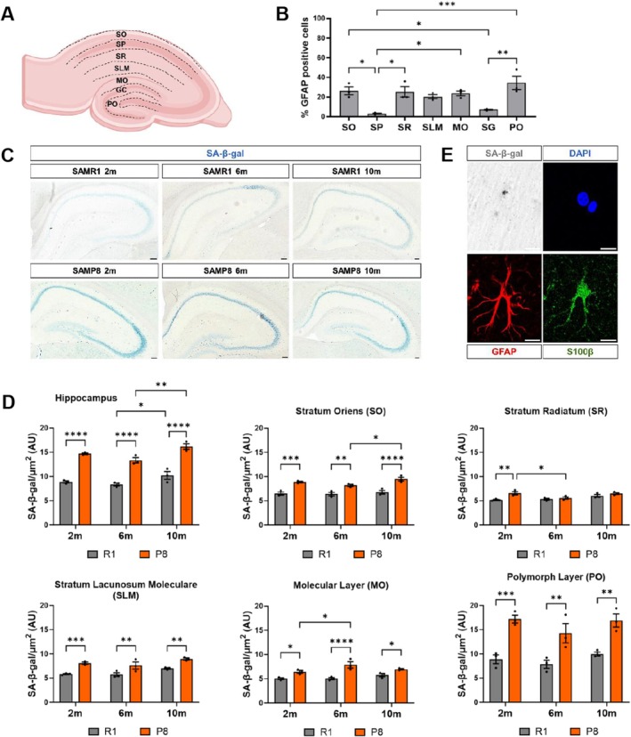
Previously, SA‐β‐gal staining has been found to increase in the hippocampus of aged rodents, being reported in pyramidal neurons of the CA3 and CA1 hippocampal regions (Geng et al. 2010; Piechota et al. 2016). However, the age‐related accumulation of senescent SA‐β‐gal+ astrocytes throughout the hippocampus has been poorly explored. To evaluate the impact of astrocyte senescence on hippocampal function, SA‐β‐gal signal intensity was measured in brain sections of 2‐, 6‐, and 10‐month‐old (2, 6, and 10‐m) SAMR1 and SAMP8 animals. We evaluated both the entire hippocampus and a selection of hippocampal layers with high astrocyte content, according to the percentage of cells expressing the astrocyte marker GFAP (Glial Fibrillary Acidic Protein) (Figure 1A,B). When comparing animals of the same age, SA‐β‐gal signal intensity in the entire hippocampus of the SAMP8 strain increased significantly compared to the SAMR1 control strain (Figure 1C,D, p < 0.0001). SA‐β‐gal signal intensity also increased from 6 to 10‐m in both strains (Figure 1D). At an early age (2‐m), all the evaluated astrocyte‐enriched hippocampal layers (Stratum Oriens–SO, Stratum Radiatum–SR, Stratum Lacunosum Moleculare–SLM, Molecular Layer–MO, and Polymorphic Layer–PO) showed signs of senescence in SAMP8 versus SAMR1. For most layers, at 6 and 10‐m SA‐β‐gal signal intensity remained higher in SAMP8. Two‐way ANOVA analysis indicated that the effect of the strain was significant for the entire hippocampus and for all the analyzed layers, with an interaction between strain and age in the SR stratum (p < 0.05). The combination of SA‐β‐gal staining with immunofluorescence detection of the mature astrocyte markers S100β (S100 calcium‐binding protein beta) and GFAP confirmed the presence of SA‐β‐gal+ astrocytes (Figures 1E and S1). Together, these data allow us to conclude that the SA‐β‐gal senescence mark is precociously increased in SAMP8 hippocampal layers with high astrocyte content compared to SAMR1 and is detected in GFAP+S100β+ astrocytes.

Senescence‐associated β‐galactosidase (SA‐β‐gal) activity in the hippocampus of SAMP8 mice shows higher intensity at 2, 6, and 10 months compared to SAMR1 mice. (A) Diagram of the hippocampal layers: Stratum oriens (SO), pyramidal layer (SP), stratum radiatum (SR), stratum lacunosum moleculare (SLM), molecular layer (MO), granule cell layer (GC) and polymorphic layer (PO). (B) Percentage of astrocytes (GFAP+) in the different hippocampal layers. (C) Representative slices of hippocampus from SAMR1 and SAMP8 mice at 2, 6, and 10 months with the SA‐β‐gal staining. (D) Quantification of the SA‐β‐gal staining intensity in the whole hippocampus and in specific layers normalized to the area (μm). (E) Representative SA‐β‐gal positive astrocyte from stratum radiatum layer with GFAP (red) and S100β (green) biomarkers. The slices have 40 μm of thickness. Three independent animals of each strain and age were analyzed ( = 3). Data are presented as mean ± SEM. Two‐way ANOVA test was performed. * < 0.05, ** < 0.01, *** < 0.001, and **** < 0.0001. Scale bar, C = 50 μm; E = 10 μm. Representation shown in A. was created with. 2 n p p p p BioRender.com
Astrocytes Derived From HippocampalNeural Stem Cells Enter Senescence and Fail to Support Synaptogenesis SAMP8
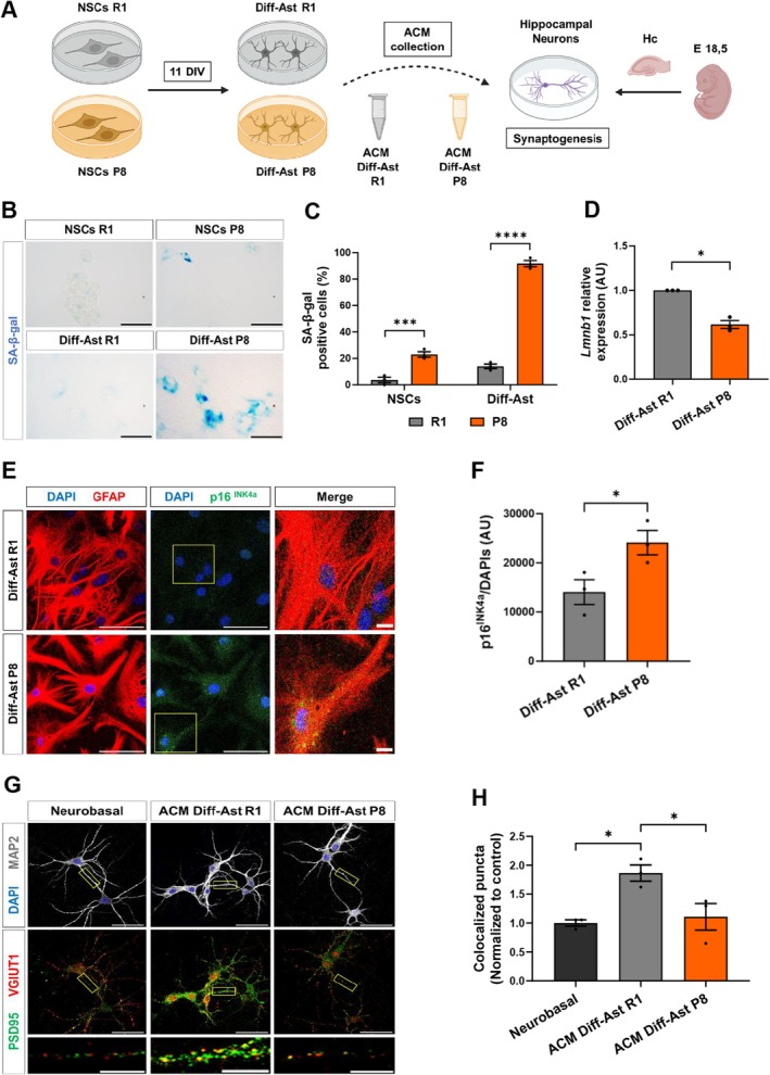
Astrocytes are key regulators of neuronal synapse formation, maintenance, and function, so we next designed experiments to explore the impact of hippocampal senescent astrocytes on hippocampal neuronal synaptogenesis. To model astrocyte senescence, we developed a simple and efficient in vitro method using hippocampal neural stem cells (NSCs) isolated from 2‐m SAMR1 and SAMP8 animals (Figure 2A). We employed Neurobasal medium supplemented with serum to stimulate astrocyte differentiation (McCarthy and De Vellis 1980). Expression of the astrocyte markers GLAST (Glutamate and aspartate transporter), ATP1B2 (ATPase Na+/K+ transporting subunit beta 2), S100β, and GFAP was monitored in the cultures (Figure S2). Quantifications showed that nearly 100% of the differentiated SAMR1 and SAMP8 cells were GFAP+S100β+ and GLAST+ATP1B2+ at 8 days in vitro (DIV), in all independent assays performed (Figure S2). Consistently, expression of the intermediate filament Nestin, a NSC and progenitor cell marker, was reduced upon differentiation (Figure S2). We also verified the acquisition of relevant astrocyte functions such as glutamate uptake, following incubation of the cells with 100 μM glutamate in HBSS. For both strains, the presence of free glutamate in the media was reduced in differentiated astrocytes compared to NSCs, indicating an increased reuptake upon differentiation (Figure S2). Thus, we concluded that astrocytes differentiated in vitro from hippocampal NSCs (Diff‐Astrocytes hereafter) acquire mature astrocytic properties.
We next compared the SA‐β‐gal senescence mark in SAMR1 and SAMP8 NSCs and Diff‐Astrocytes and quantified the percentage of positive cells. As expected, SAMP8 NSC and SAMP8 Diff‐Astrocyte cultures showed a higher proportion of SA‐β‐gal+ cells compared to control SAMR1 cultures (p < 0.0001; Figure 2B,C). Interestingly, SAMP8 NSCs already displayed this senescence mark compared to SAMR1 NSCs, but the difference was greatly exacerbated upon NSC differentiation into astrocytes. Reduced Lamin B1 expression (p < 0.05; Figure 2D) and increased p16INK4a protein levels (p < 0.05; Figure 2E,F), two other hallmarks of cellular senescence (Matias et al. 2021), were also detected in SAMP8 Diff‐Astrocytes compared to SAMR1 Diff‐Astrocytes by RT‐qPCR and immunofluorescence, respectively. Expression of Cdkn1a/p21CIP1 and SASP genes further supported the acquisition of senescence (Figure S2).
Next, we evaluated if senescence influenced the synaptogenic capacity of the Diff‐Astrocyte cultures. To this end, we established primary hippocampal neuronal cultures from wild‐type embryos (E18.5) and tested the effect of astrocyte conditioned media (ACM) collected from SAMR1 and SAMP8 Diff‐Astrocytes (Figure 2A). Excitatory synapse formation was evaluated based on the immunofluorescence and co‐localization image analysis of the pre‐synaptic vesicle marker VGLUT1 (Vesicular Glutamate Transporter 1) and the post‐synaptic marker PSD95 (Post‐Synaptic Density protein 95), as previously described (Matias et al. 2021). ACM collected from SAMR1 Diff‐Astrocytes promoted co‐localization of the pre‐ and post‐synaptic markers, demonstrating a positive impact on the generation of new synapses compared to control medium (p < 0.05; Figure 2G,H). Most importantly, ACM from SAMP8 Diff‐Astrocyte cultures had no effect, demonstrating a loss of synaptogenic function in senescent astrocytes. Potential neurotoxic effects did not appear to drive the synaptic density changes (Figure S3A). Altogether, these results indicate that astrocytes differentiated in vitro from hippocampal SAMP8 NSCs enter senescence (as shown by the rise in SA‐β‐gal, p16INK4a and p21CIP1, and the reduction in Lamin B1) and are deficient in promoting synaptogenesis compared to control (SAMR1) astrocytes, providing a suitable model to study the impact of astrocyte senescence in astrocyte‐neuron communication.

Differentiated astrocytes (Diff‐Ast) derived from SAMP8 neural stem cells display hallmarks of senescence and synaptogenic dysfunction. (A) Schematic diagram of NSCs culture and differentiation to astrocytes. (B, C) SA‐β‐gal activity in NSCs and Diff‐Ast SAMR1 (R1) and SAMP8 (P8) cultures. (D) RT‐qPCR of( = 0.013) in Diff‐Ast SAMR1 and SAMP8 cultures. (E, F) Immunostaining of GFAP (red), p16INK4a (green), and p16INK4a quantification intensity in Diff‐Ast SAMR1 and SAMP8 cultures. (G, H) Immunostaining of MAP2 (gray), PSD95 (green) and VGLUT1 (red), and quantification of excitatory pre‐ (VGLUT1) and postsynaptic (PSD95) vesicles colocalization in hippocampal neurons treated with ACMs from Diff‐Ast SAMR1 and SAMP8 cultures. Three independent experiments were analyzed per mouse strain and cell type ( = 3). Data are presented as mean ± SEM. One‐way ANOVA Tukey's multiple comparisons test was performed in (C) and (H). One‐sample‐test was done in (D). Unpaired‐test in (F). * < 0.05, ** < 0.01, *** < 0.001, and **** < 0.0001. Scale bar: B, E, and G = 50 μm; G = 10 μm (representative neurite). Representation shown in (A) was created with. Lmnb1 p n t t p p p p BioRender.com
Acutely Isolated‐2Hippocampal Astrocytes FromAnimals Exhibit Senescence Marks and Fail to Support Synaptogenesis ACSA SAMP8 +
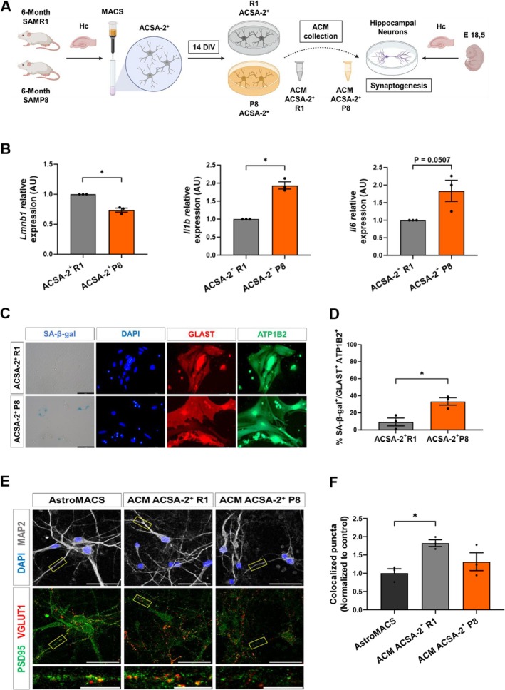
To further corroborate these findings, we next isolated astrocytes directly from the adult hippocampus of 6‐m SAMR1 and SAMP8 mice employing magnetic cell separation (MACS), taking advantage of the astrocyte cell surface marker ACSA‐2 (encoded by Atp1b2) (Figure 3A). We analyzed the acutely isolated ACSA‐2+ cells by RT‐qPCR for senescence and SASP markers. Lamin B1 expression was significantly decreased (p < 0.05; Figure 3B), while IL‐1β expression was upregulated (p < 0.05; Figure 3B) in ACSA‐2+ SAMP8 cells compared to ACSA‐2+ SAMR1 cells. Hippocampal ACSA‐2+ cells were then cultured in vitro in defined astrocytic growth medium (without serum) and were analyzed by immunocytochemistry, with specific astrocyte markers and SA‐β‐gal staining (Figure 3C). The vast majority of the hippocampal ACSA‐2+ isolated cells expressed astrocyte markers upon culture and were double‐positive for GLAST and ATP1B2 (average GLAST+ATP1B2+ cells ± S.E.M: 96.7% ± 0.7% in SAMR1 cultures and 93.3% ± 2.0% in SAMP8 cultures, n = 3). On average, 9.3% ± 4.5% of the GLAST+ATP1B2+ astrocytes were SA‐β‐gal+ in SAMR1 primary astrocyte cultures, and this percentage raised 3‐fold in SAMP8 primary astrocyte cultures (33.2% ± 4.1%, p < 0.05; Figure 3C,D). Similar results were obtained when SAMR1 and SAMP8 astrocytes were isolated and cultured from 2 and 10‐m animals (Figure S1).
Next, we evaluated the synaptogenic activity of ACM collected from primary ACSA‐2+ astrocytes, based on the co‐localization of VGLUT1 and PSD95 in ACM‐treated hippocampal neuronal cultures (Figure 3E,F). SAMR1 ACSA‐2+ ACM significantly promoted synapse formation compared to non‐conditioned control medium (p < 0.05; Figure 3F). In contrast, no synaptogenic effect was found when neurons were treated with SAMP8 ACSA‐2+ ACM (Figure 3F). One‐way ANOVA revealed no significant differences in neuronal density across conditions, suggesting that neurotoxic effects were not the primary driver of the observed reduction in synaptic density in the senescent SAMP8 medium (Figure S3B).
In summary, these results show that ACSA‐2+ hippocampal astrocytes isolated from SAMP8 animals show senescence marks and are deficient in synaptogenic activity. While primary SAMR1 hippocampal astrocytes release factors into the ACM that promote the formation of new excitatory glutamatergic synapses in hippocampal neuronal cultures, this capacity is lost in primary SAMP8 hippocampal astrocytes, in line with the results obtained with senescent astrocytes differentiated from SAMP8 NSCs.

6‐month‐old ACSA‐2 SAMP8 primary cultured astrocytes display signs of senescence and deficiencies in synaptogenic activity. (A) Experimental design of ACSA‐2+ astrocytes isolation from the 6‐month‐old SAMR1 and SAMP8 hippocampus. (B) RT‐qPCR of( = 0.016),( = 0.011) and( = 0.050) in ACSA‐2+ SAMR1 and SAMP8 cells isolated from 6‐m mice. (C, D) Representative images and quantification of SA‐β‐gal activity in ACSA‐2+ primary cultures with astroglial GLAST (red) and ATP1B2 (green) markers after 14 days in culture. (E, F) Immunostaining of MAP2 (gray), PSD95 (green) and VGLUT1 (red), and quantification of excitatory pre‐ (VGLUT1) and post‐synaptic (PSD95) vesicle co‐localization in hippocampal neurons treated with ACMs from ACSA‐2+ SAMR1 and SAMP8 astrocytes of 6‐m. Three independent experiments were analyzed for each mouse strain ( = 3). Data are presented as mean ± SEM. One‐sample‐test was performed in (C). Unpaired‐test was done in (D). One‐way ANOVA test was performed in (F). * < 0.05. Scale bar: 50 and 10 μm. Representation shown in (A) was created with. Lmnb1 p Il1β p Il6 p n t t p BioRender.com
Thrombospondin‐1 Expression Is Downregulated in Senescent Hippocampal‐2Astrocytes and in Senescent Astrocytes Derived FromNeural Stem Cells SAMP8 ACSA SAMP8 +
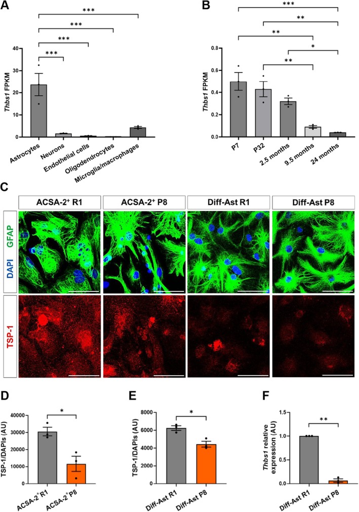
We then followed a candidate approach to uncover the identity of putative factors repressed in hippocampal senescent astrocytes that could account for the loss of synaptogenic function. Healthy brain astrocytes release molecules to the extracellular space that contribute to the formation of synapses (Ullian et al. 2004). Thrombospondin‐1 (TSP‐1, encoded by the Thbs1 gene) has emerged as an astrocyte‐secreted protein with prominent effects on synaptogenesis and neuritogenesis during development (Cheng et al. 2016; Ikeda et al. 2010; Yu et al. 2008). We explored available bulk RNAseq databases in silico and confirmed the enriched expression of Thbs1 in astrocytes among all other major cell types of the murine brain (Zhang et al. 2014) (Figure 4A). Inspection of an RNAseq dataset from hippocampal astrocytes of postnatal, young, middle‐age, and old wild‐type animals highlighted the downregulation of Thbs1 expression during aging (Clarke et al. 2018) (Figure 4B). Given Thbs1 gene expression is also reduced in astrocytes isolated from other brain areas during aging (Boisvert et al. 2018), we decided to delve deeper into Thbs1/TSP‐1 expression in senescent astrocytes.
We evaluated Thbs1 mRNA and TSP‐1 protein levels in senescent SAMP8 hippocampal astrocytes by RT‐qPCR and immunocytochemistry. Our results demonstrate that primary SAMP8 ACSA‐2+ astrocytes isolated from the hippocampus show significantly lower TSP‐1 protein levels compared to SAMR1 ACSA‐2+ astrocytes (p < 0.05; Figure 4C,D). On the other hand, SAMP8 Diff‐Astrocyte cultures also show decreased TSP‐1 protein levels, as quantified by immunofluorescence (p < 0.05; Figure 4C,E) and Western blot (Figure S3), and reduced Thbs1 gene expression compared to SAMR1 Diff‐Astrocytes (p < 0.01; Figure 4F).
![6‐month‐old ACSA‐2+ SAMP8 primary cultured astrocytes and differentiated astrocytes derived from SAMP8 neural stem cells display loss in TSP‐1 levels. (A) RNA‐seq transcriptional profile ofgene in astrocytes, neurons, endothelial cells, oligodendrocytes and microglia in P7 (postnatal day) mice. (B) RNA‐seq transcriptional profile ofgene at different stages of development and aging in astrocytes from mouse hippocampus. (C) Representative images of GFAP (green) and TSP‐1 (red) immunostaining in ACSA‐2+ primary astrocytes cultures of the SAMP8 and SAMR1 strains (14 DIV) and in Diff‐Ast SAMP8 and SAMR1 strains (11 DIV). (D, E) Quantification of TSP‐1 protein shows lower levels in SAMP8 ACSA‐2+ astrocytes cultures ( = 0.021) and in Diff‐Ast SAMP8 ( = 0.011) than in the control SAMR1 strain. (F) RT‐qPCR of( = 0.001) demonstrates lower gene expression levels in Diff‐Ast SAMP8 compared to the control line SAMR1. Three independent experiments per cell type were analyzed ( = 3). Data are presented as mean ± SEM. Unpaired‐test was performed in (D, E). One‐sample‐test was done in (F). * < 0.05, ** < 0.01, *** < 0.001. Scale bar: 50 μm. Data in (A) and (B) were obtained from databases produced by the Barres lab (Zhang et al. () and Clarke et al. () available at). Thbs1 Thbs1 p p Thbs1 p n t t p p p [2014] [2018] https://brainrnaseq.org/](https://europepmc.org/articles/PMC12813271/bin/ACEL-25-e70382-g007.jpg "Click to view full size")
6‐month‐old ACSA‐2+ SAMP8 primary cultured astrocytes and differentiated astrocytes derived from SAMP8 neural stem cells display loss in TSP‐1 levels. (A) RNA‐seq transcriptional profile ofgene in astrocytes, neurons, endothelial cells, oligodendrocytes and microglia in P7 (postnatal day) mice. (B) RNA‐seq transcriptional profile ofgene at different stages of development and aging in astrocytes from mouse hippocampus. (C) Representative images of GFAP (green) and TSP‐1 (red) immunostaining in ACSA‐2+ primary astrocytes cultures of the SAMP8 and SAMR1 strains (14 DIV) and in Diff‐Ast SAMP8 and SAMR1 strains (11 DIV). (D, E) Quantification of TSP‐1 protein shows lower levels in SAMP8 ACSA‐2+ astrocytes cultures ( = 0.021) and in Diff‐Ast SAMP8 ( = 0.011) than in the control SAMR1 strain. (F) RT‐qPCR of( = 0.001) demonstrates lower gene expression levels in Diff‐Ast SAMP8 compared to the control line SAMR1. Three independent experiments per cell type were analyzed ( = 3). Data are presented as mean ± SEM. Unpaired‐test was performed in (D, E). One‐sample‐test was done in (F). * < 0.05, ** < 0.01, *** < 0.001. Scale bar: 50 μm. Data in (A) and (B) were obtained from databases produced by the Barres lab (Zhang et al. () and Clarke et al. () available at). Thbs1 Thbs1 p p Thbs1 p n t t p p p [2014] [2018] https://brainrnaseq.org/
Thrombospondin‐1 Expression and Excitatory Synapses Decrease in theHippocampus SAMP8
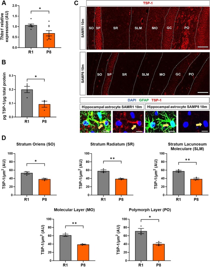
In order to further explore Thbs1/TSP‐1 expression in the SAMP8 hippocampus in vivo, we measured Thbs1 mRNA levels by RT‐qPCR (Figure 5A) and total TSP‐1 protein levels by ELISA (Figure 5B) in hippocampal tissue extracts from 10‐m animals. Results confirmed the reduction in total Thbs1 mRNA and TSP‐1 protein content in SAMP8 hippocampus relative to SAMR1. This was further validated by immunofluorescence in hippocampal sections from SAMP8 relative to SAMR1 animals (Figure 5C), where TSP‐1 signal was found both co‐localizing with the astrocytic marker GFAP and in the extracellular matrix surrounding the astrocytic processes. Confocal image analysis showed decreased TSP‐1 levels in most astrocyte‐enriched layers of the 10‐m SAMP8 hippocampus compared to SAMR1 (Figure 5D). Finally, we also analyzed VGLUT1 and PSD95 co‐localization in 10‐m SAMP8 and SAMR1 hippocampal sections. As shown in Figure S4, we detected a significant decrease in co‐localized VGLUT1 and PSD95 excitatory synaptic puncta in most analyzed SAMP8 hippocampal layers, and a significant correlation between TSP‐1 signal intensity and puncta co‐localization (Figure S4). These data support the notion that loss of TSP‐1 expression in hippocampal senescent astrocytes may contribute to the reduction in excitatory synapses in SAMP8 animals.

The hippocampus of 10‐month‐old SAMP8 mice shows deficiencies in TSP‐1 levels. (A) RT‐qPCR of( = 0.022) in SAMR1 ( = 7 independent animals) and SAMP8 ( = 8 independent animals). (B) TSP‐1 quantification by ELISA test in 10‐m SAMR1 ( = 4 independent animals) and SAMP8 ( = 3 independent animals) mice normalized to total protein levels. (C, D) Immunohistochemical analyses and TSP‐1 intensity quantification in 10‐m SAMR1 and SAMP8 hippocampus are shown. Representative astrocytes from the SLM layer in the hippocampus using GFAP (green) as an astroglial marker, and TSP‐1 (red) are depicted magnified. TSP‐1 signal intensity quantification was performed in each layer of the hippocampus and normalized to the quantification area (μm2). Three independent animals of each strain were analyzed for immunohistochemical analysis ( = 3). Data are presented as mean ± SEM. Unpaired‐test was performed. * < 0.05 and ** < 0.01. Scale bar: 100 μm; 10 μm (representative astrocyte). Thbs1 p n n n n n t p p
‐1 Released by HealthyAstrocytes Is a Major Contributor to Excitatory Synaptogenesis Through α2δ‐1 Receptor Signaling TSP SAMR1
We next designed a functional assay to explore whether the synaptogenic effect induced by SAMR1 astrocytes is related to TSP‐1 secretion. For this purpose, we employed Gabapentin (GBP), a pharmacological antagonist that blocks TSP‐1 signaling through its neuronal receptor, the voltage‐gated calcium channel (VGCC) subunit α2δ‐1 (Cheng et al. 2016; Eroglu et al. 2009). The α2δ‐1 receptor is encoded by the Cacna2d1 gene, which is highly expressed in neurons throughout the CNS. The α2δ‐1 receptor is enriched in hippocampal pyramidal neurons (Cole et al. 2005), and as shown by RT‐qPCR, is expressed in the hippocampal neurons used in our cell culture assays (Figure S5). As shown in Figure 6, GBP completely blocked the pro‐synaptogenic effect of SAMR1 Diff‐Astrocyte ACM (p < 0.01; Figure 6A,C). Similarly, GBP inhibited the positive effect of the SAMR1 ACSA‐2+ ACM on the formation of new synapses (p < 0.05; Figure 6B,D). No significant differences were found in GBP‐treated cultures exposed to SAMP8 Diff‐Astrocyte ACM or SAMP8 ACSA‐2+ ACM.
In summary, these results allow us to establish that the positive effect on the formation of new excitatory synapses triggered by healthy SAMR1 Diff‐Astrocytes and SAMR1 ACSA‐2+ primary hippocampal astrocytes is related to the presence of high TSP‐1 levels in the ACM, which signals through the neuronal receptor α2δ‐1.

The competitive TSP‐1 receptor antagonist gabapentin (GBP) blocks the synaptogenic effect of SAMR1 ACMs. (A and C) Immunostaining of MAP2 (gray), VGLUT1 (red) and PSD95 (green), and quantification of excitatory pre‐ (VGLUT1) and postsynaptic (PSD95) vesicles colocalization in hippocampal neurons treated with ACMs from Diff‐Ast SAMR1 and SAMP8 with or without GBP 32 μM. (B and D) Immunostaining of MAP2 (gray), VGLUT1 (red) and PSD95 (green), and quantification of excitatory pre‐ (VGLUT1) and postsynaptic (PSD95) colocalization vesicles in hippocampal neurons treated with ACMs from ACSA‐2SAMR1 and SAMP8 of 6‐m mice with or without GBP 32 μM. Three independent experiments were analyzed per cell type and experimental condition. Data are presented as mean ± SEM. One‐way ANOVA Tukey's multiple comparisons test was performed. * < 0.05, ** < 0.01, and *** < 0.001. Scale bar: 50 μm. + p p p
‐1 Supplementation Recovers the Synaptogenic Activity of Senescent Astrocyte Conditioned Medium TSP
Finally, we explored whether in vitro treatment with purified recombinant TSP‐1 restored the loss of synaptogenic activity of senescent SAMP8 astrocytes. Supplementing SAMP8 Diff‐Astrocyte ACM with TSP‐1 was sufficient to recover glutamatergic excitatory synapse formation in primary hippocampal neuronal cultures (p < 0.05; Figure 7A,B). As a control, we further demonstrated that the pharmacological antagonist GBP blocked the TSP‐1 effect on the co‐localization of VGLUT1 and PSD95 pre‐ and post‐synaptic markers (Figure S5). Overexpression of the mouse Thbs1 gene in SAMP8 differentiated astrocytes (Figure S5), followed by treatment of neuronal cultures with the resulting conditioned medium (Figure 7C,D) showed that restoring TSP‐1 expression suffices to rescue synaptogenic activity in senescent SAMP8 astrocytes.

TSP‐1 rescues the synaptogenic function of Diff‐Ast SAMP8 ACM. (A, B) Immunostaining of MAP2 (gray), VGLUT1 (red) and PSD95 (green), and quantification of excitatory pre‐ (VGLUT1) and postsynaptic (PSD95) vesicles colocalization in hippocampal neurons treated with ACM from Diff‐Ast SAMP8 with or without TSP‐1 (250 ng/mL) supplementation. (C, D) Immunostaining of MAP2 (gray), VGLUT1 (red) and PSD95 (green), and quantification of excitatory pre‐ (VGLUT1) and postsynaptic (PSD95) vesicles colocalization in hippocampal neurons treated with ACM from transfected Diff‐Ast SAMP8 overexpressingand GFP, or pcDNA3 and GFP as a control. Three independent experiments were analyzed per cell type and experimental condition. Data are presented as mean ± SEM. One‐way ANOVA Tukey's multiple comparisons test was performed. * < 0.05 and ** < 0.01. Scale bar: 50 μm. mThbs1 p p
Discussion
In this study, we employed the accelerated aging mouse model SAMP8 to characterize the accumulation of senescent astrocytes in the aged hippocampus and study the functional impact of senescence on astrocyte‐neuron communication and excitatory synapse formation. Astrocytes have a variety of biological functions, including the regulation of new synapse formation (Verkhratsky and Nedergaard 2018). This function is mainly mediated by astrocyte‐secreted protein factors (Baldwin and Eroglu 2017). Astrocytes regulate the formation of different types of synapses, such as glutamatergic (Ullian et al. 2001), GABAergic (Elmariah et al. 2005; Hughes et al. 2010), glycinergic (Cuevas et al. 2005), and cholinergic synapses (Reddy et al. 2003; Cao and Ko 2007). Here, we confirmed the accumulation of SA‐β‐gal+ hippocampal astrocytes in SAMP8 in vivo, both in histological sections and in isolated astrocytes expressing the surface antigen ACSA‐2. The raise in senescent astrocytes was accompanied by a reduction at 10 months of age in the co‐localization of the pre‐ and post‐synaptic excitatory glutamatergic markers, VGLUT1 and PSD95, a proxy of synapse number. Using astrocyte cultures and ACMs, we focused on evaluating the impact of the senescent astrocyte secretome on synapse formation. We uncovered a reduction in TSP‐1, an important astrocyte‐secreted protein involved in the regulation of spine development and synaptogenesis, in senescent astrocytes. We showed that addition of exogenous TSP‐1, or overexpression of Thbs1, is sufficient to rescue the synaptogenic deficiency of senescent astrocytes.
Previous studies showed that senescence can be induced in vitro in murine and human astrocytes, through long‐term cell culture (Geng et al. 2010; Kawano et al. 2012; Matias et al. 2021; Saenz‐Antoñanzas et al. 2024), oxidative stress treatment (Simmnacher et al. 2020), or X‐irradiation (Limbad et al. 2020). Mimicking astrocyte aging in vitro through prolonged cell culture increases reactive oxygen species production and compromises the astrocyte's neuroprotective capacity (Pertusa et al. 2007). A loss of synaptogenic function has been reported in cortical neonatal astrocytes from wild‐type mice that entered senescence after several weeks of in vitro cell culture, but the mechanism underlying this phenotype has remained elusive (Kawano et al. 2012; Matias et al. 2021). To our knowledge, our study is the first to demonstrate that synaptogenic defects characterize naturally senescent astrocytes directly purified from the aged brain, shedding light on the molecular basis of their lack of synaptogenic function and linking TSP‐1 loss to astrocyte senescence.
For the isolation of senescent astrocytes from adult hippocampal tissue, we took advantage of the positive selection with antibodies against the astroglial surface antigen ACSA‐2. Primary ACSA‐2+ astrocyte cultures were established and maintained for 14 DIV in a defined serum‐free medium. Despite the low yield of this type of culture, the approach is of great interest for studying the properties of astrocytes ex vivo, since in classical methodologies based on postnatal primary cultures expanded in the presence of serum, the characteristics of the astrocytes change and a reactive glial phenotype is induced (Caldwell et al. 2022). We found no significant differences in the purity of the ACSA‐2 cultures between strains, with the astroglial markers GLAST and ATP1B2 reaching values close to 95% of positive cells in all cases. SA‐β‐gal staining demonstrated a higher proportion of senescent GLAST+ ATP1B2+ SA‐β‐gal+ astrocytes in SAMP8 cultures compared to SAMR1 cultures. We also confirmed other senescence markers in ACSA‐2+ SAMP8 astrocytes by RT‐qPCR (decreased expression of the gene encoding Lamin B1 and increased expression of the gene encoding IL‐1β). Thus, this strategy allows to obtain naturally senescent astrocytes directly from the aged brain. Most importantly, it allowed us to prove that the glutamatergic synaptogenic function of primary senescent astrocytes is greatly impaired, as shown by the lack of effect of the ACSA‐2+ SAMP8 ACM on wild type murine hippocampal primary neuronal cultures. Results were also confirmed when using ACM collected from in vitro differentiated SAMP8 astrocytes.
On the other hand, we derived SAMR1 and SAMP8 Diff‐Astrocytes from hippocampal NSCs. This complementary strategy represents a simple and practical methodology for studying astrocyte senescence, allowing for large‐scale preparations without the need to promote the acquisition of senescence through long‐term culture or exposure to cell damaging agents. This approach is very efficient and reproducible, generating SAMP8 astrocyte monolayers highly enriched (~90%) in SA‐β‐gal+ senescent astrocytes, with low Lamin B1 expression and elevated levels of p16INK4a. The rise in the cell cycle inhibitor p16INK4a has been widely reported as a senescence hallmark (López‐Otín et al. 2023). Transgenic mice have been developed to visualize and eliminate senescent cells from aged tissues based on this marker (p16‐INK‐ATTAC mice, Baker et al. 2011; p16‐3MR mice, Demaria et al. 2014). Astrocytes derived from postnatal mice show higher levels of p16INK4a after 30–35 DIV (Matias et al. 2021). In post‐mortem brains of Parkinson's disease patients, astrocytes accumulate senescence marks and p16INK4a is increased in the substantia nigra (Chinta et al. 2018). Here we report that astrocytes differentiated from SAMP8 NSCs show higher p16INK4a content compared to SAMR1 astrocytes after 11 DIV. Along the same lines, mRNA levels of other senescence markers corroborate the senescent state of SAMP8 Diff‐Astrocytes. We detected a significant decrease in Lmnb1 expression (encoding Lamin B1), a significant increase in Cdkn1a expression (encoding p21CIP1), and a trend towards the upregulation of the genes that code for IL‐1β and IL‐6, two secreted pro‐inflammatory molecules related to SASP. Decreased Lamin B1 and increased IL‐1β expression were confirmed in SAMP8 ACSA‐2+ astrocytes. Interestingly, decreased Lamin B1 content has been proposed as a hallmark of astrocyte senescence in the hippocampal dentate gyrus of aged mice (Matias et al. 2021). Furthermore, a general reduction in Lamin B1 has been reported in the hippocampal granule cell layer of post‐mortem human tissue from elderly individuals. In the same tissue samples, hippocampal astrocytes show reduced nuclear circularity, possibly related to the loss of Lamin B1 (Matias et al. 2021).
Studying the molecular basis of astrocyte‐neuron communication requires analyzing the protein composition of the astrocyte secretome. Leading groups in the field have demonstrated the poor coincidence between changes in astrocyte gene expression and secretome (Caldwell et al. 2022), so astrocyte transcriptomics are not informative enough if changes are not confirmed at the protein level. We focused on evaluating the synaptogenic effect of the protein factors secreted by healthy astrocytes and their alteration in senescent astrocytes. We observed that, for the formation of new excitatory glutamatergic synapses, SAMR1 control ACM is pro‐synaptogenic, as expected based on the literature (Baldwin and Eroglu 2017), while SAMP8 senescent ACM shows a loss of function. TSP‐1 protein levels were significantly reduced in astrocytes from the SAMP8 strain compared to SAMR1. TSP‐1 is a secreted matricellular protein from the thrombospondin family, described as a key factor in the regulation of structural synapse formation in several neuronal types (Christopherson et al. 2005; Eroglu et al. 2009; Xu et al. 2010; Cheng et al. 2016). TSP‐1 and TSP‐2 deficient mice have lower synaptic density in the cortex (Christopherson et al. 2005), so a similar phenotype could be expected in the hippocampus, taking into account that the α2δ‐1 receptor is enriched in both cortical and hippocampal pyramidal neurons (Cole et al. 2005). Furthermore, α2δ‐1 knockout animals show a significant reduction in the number, degree of maturation, and activity of excitatory synapses in the cortex (Risher et al. 2018), again highlighting the relevance of TSP‐1/α2δ‐1 signaling for synaptogenesis.
To support the role of hippocampal astrocyte‐released TSP‐1, we performed a functional assay using gabapentin (GBP), a drug used to treat epilepsy and neuropathic pain that acts as an antagonist competitor of the TSP‐1 receptor α2δ‐1 in neurons (Eroglu et al. 2009; Cheng et al. 2016). The synaptogenic activity of thrombospondin is mediated by its EGF‐like domain, which binds to the extracellular domain of α2δ‐1. The interaction is thought to cause a conformational change in α2δ‐1 that in turn allows the recruitment of a synaptogenic signaling complex (Baldwin and Eroglu 2017). At the post‐synaptic level, this complex regulates synaptogenesis through the Rho GTPase Rac1, which in turn promotes the reorganization of the actin cytoskeleton (Risher et al. 2018). Our results clearly show that GBP blocks excitatory synapse formation in hippocampal neurons when added to the SAMR1 ACM.
TSP‐1 promotes the formation of silent synapses (Christopherson et al. 2005), which contain NMDA receptors but lack functional AMPA receptors. These synapses are inactive under baseline conditions but can be rapidly "unsilenced" during activity‐dependent processes such as long‐term potentiation (LTP) (Malinow and Malenka 2002; Kerchner and Nicoll 2008). "Unsilencing" occurs via the insertion of AMPA receptors, thereby strengthening synaptic transmission and facilitating learning and memory. Therefore, silent synapses provide a reservoir of modifiable connections that act as a versatile substrate for experience‐dependent synaptic plasticity.
Previous work shows that post‐synaptically silent synapses persist in the hippocampus of both adult and aged rats (Sametsky et al. 2010). Although their prevalence declines slightly with age—potentially contributing to age‐related cognitive impairment—they remain a viable target for therapeutic interventions aimed at enhancing cognitive function in aging (Kumar et al. 2007; Sametsky et al. 2010). Age‐related cognitive decline has been attributed to dysregulated activity‐dependent plasticity at functional synapses and/or impaired recruitment of AMPA receptors into silent synapses (Sametsky et al. 2010). Reduced TSP1 production in senescent animals may limit the available pool of silent synapses—at least in the accelerated aging SAMP8 mouse model—thereby possibly contributing to cognitive decline.
TSP‐1 expression alterations have been previously related to synaptic defects in several pathologies, but the contribution of senescent astrocytes to these syndromes remains underexplored. Alzheimer's disease (AD) is a devastating age‐related neurodegenerative disorder that is associated with massive synapse loss in the early clinical phase (Masliah et al. 1991; Sze et al. 1997). TSP‐1 expression is decreased in neurons of AD patients (Buee et al. 1992). Interestingly, thrombospondins accumulate in Ab plaques of post‐mortem AD patient brain samples compared to control cases. Moreover, it has been suggested that increased thrombospondin expression in human brain evolution may contribute to the cognitive abilities of the human species compared to chimpanzees and macaques, but also to the increased human vulnerability to AD and neurodegeneration (Cáceres et al. 2007). Loss of thrombospondin expression has been reported as well in astrocytes from Down's syndrome, a disease associated with a reduction in synaptic density (Garcia et al. 2010). Abnormal hippocampal synaptogenesis also characterizes Fragile X Syndrome (FXS), the major cause of inherited mental retardation (Jawaid et al. 2018). Interestingly, astrocytes isolated from a mouse model of FXS display loss of TSP‐1 protein expression. Alterations in excitatory synapse formation of FXS hippocampal neurons are prevented in the presence of healthy ACM or following the administration of TSP‐1 (Cheng et al. 2016). Therefore, soluble TSP‐1 has been proposed as a potential therapeutic target for mental retardation syndromes. Our results extend its potential use to treat age‐related conditions that involve synaptic decline.
In summary, our results highlight the contribution of astrocyte senescence to neuronal microenvironment dysfunction during aging and, in particular, reveal a potential role in hippocampal synapse reduction. Our study also reinforces the use of the SAMP8 strain for the study of brain aging and provides two reproducible protocols for obtaining senescent astrocytes in vitro. Combining these methodologies, we demonstrate that TSP‐1 released by healthy adult hippocampal astrocytes is a major contributor to synapse formation through α2δ‐1 receptor signaling in neurons, and that reduced TSP‐1 expression in senescent astrocytes impairs their synaptogenic capacity. Brain repair mechanisms aiming to recover synapse loss may take into account the poor performance of senescent astrocytes. Our findings also reinforce the use of senotherapeutics as promising strategies to mitigate the effects of aging on brain function.
Author Contributions
S.E. and H.M. designed research; S.E., L.C.‐C., and E.J.‐E. performed research and analyzed data; S.E. and H.M. wrote the first draft of the manuscript. All authors approved the manuscript.
Funding
This work was supported by a predoctoral fellowship from the Ministry of Science, Technology, Knowledge and Innovation of Chile to S.E. and grants PID2022‐141707NBI00 from the Spanish Ministry of Science and Innovation and CIAICO/2022/74 from the Generalitat Valenciana to H.M.
Conflicts of Interest
The authors declare no conflicts of interest.