
What this is
- Osteoarthritis (OA) affects 595 million people globally, with limited treatment options.
- Cell-free therapy using () shows potential for cartilage repair.
- This examines trends in EV-based therapies for OA from 1991 to 2024.
Essence
- The analysis reveals a rapid increase in publications on EV-based cell-free therapy for OA, particularly since 2019, with China leading in research output and citations.
Key takeaways
- China produced the highest number of publications (202) and citations (6063) in EV-based OA research, indicating its dominance in this field.
- The International Journal of Molecular Sciences published the most articles (26), while Biomaterials received the highest citations, showcasing key journals in this research area.
- Research themes include therapeutic mechanisms of in OA, advances in cell-free treatments, and exosome engineering, reflecting a shift towards innovative therapeutic strategies.
Caveats
- The study identifies a lack of international collaboration, with most research being domestic, which may limit the diversity of insights in EV-based OA therapies.
- Despite the growth in publications, long-term safety and efficacy of EV therapies in clinical settings remain to be thoroughly evaluated.
Definitions
- extracellular vesicles (EVs): Nanoparticles secreted by cells, containing proteins, lipids, and nucleic acids that facilitate intercellular communication.
- bibliometric analysis: A quantitative method for assessing publication patterns, citation networks, and research trends in scientific literature.
Simplified
Introduction
The global healthcare landscape in the twenty-first century faces unprecedented challenges from chronic degenerative conditions, among which osteoarthritis (OA) emerges as a particularly prominent concern [1, 2]. In 2020, OA affected 595 million people, representing 7.6% of the global population. Over three decades, the incidence has surged by 132.2%, with projections indicating a further 60–100% increase by 2050 [3 –5]. The major risk factors include age, female gender, excessive weight, and previous joint injuries [6]. Marked by deteriorating cartilage, hardened subchondral bone, and bone spurs, OA stands as the primary cause of mobility restrictions in adults and skeletal deformities in older populations [5, 7, 8]. Despite numerous early-stage treatments—from medications and injections to bracing and lifestyle changes—therapeutic outcomes remain suboptimal [9]. Failed conservative management necessitates surgical interventions like arthroscopic repair or joint replacement, which involve significant trauma and prosthetic limitations [10, 11]. These procedures also carry complication risks and generate substantial direct healthcare costs and indirect economic losses through reduced workforce participation [12, 13].
Recent research, including both animal studies and clinical trials, has validates mesenchymal stem cells as an efficacious treatment for cartilage injuries and osteoarthritis [14, 15]. The clinical application of mesenchymal stem cells is limited by rigorous protocols for harvesting and transportation, coupled with potential risks of infection and neoplasia [16 –18]. With advancing tissue engineering research, studies reveal that mesenchymal stem cells function through paracrine effects, with their secreted extracellular vesicles modulating the intra-articular microenvironment and regulating cellular migration and proliferation [19, 20]. Moreover, due to their cell-free nature, these vesicles possess advantages including non-immunogenicity, non-tumorigenicity, and ease of storage and transportation [21 –24]. Membrane-bound nanoparticles known as extracellular vesicles are secreted by cells and found throughout biological fluids. These structures facilitate intercellular signaling by conveying bioactive cargo—including proteins, lipids, and various nucleic acids (mRNA, miRNA, DNA)—thereby orchestrating physiological and pathological processes [22, 25]. Based on diameter and biogenesis mechanisms, they are classified into the exosomes, microvesicles, and apoptotic bodies [26]. Among various EVs subtypes, exosomes represent the most extensively studied population and have been shown to regulate osteoarthritic cartilage extracellular matrix metabolism, inflammatory responses, and angiogenesis. Their bioactive cargo, including proteins and nucleic acids, influences OA progression through paracrine or endocrine mechanisms targeting specific cells [27 –29]. Research also indicates that microvesicles, like exosomes, can regulate OA chondrocyte metabolism and exhibit anti-inflammatory and chondroprotective effects, counteracting IL-1β's influence. MVs exhibit more pronounced effects, possibly due to Annexin A1 involvement, offering novel therapeutic strategies for joint diseases [30 –34]. In the context of OA, EVs-based approaches have manifested particular promise, offering potential solutions for both disease modification and tissue regeneration [30, 35]. The scientific community's growing interest in the applications of EVs for OA treatment has catalyzed an unprecedented surge in research activities [23]. This expansion spans multiple scientific domains, encompassing fundamental biological investigations, innovation in organizational engineering [21], mechanistic studies of therapeutic effects [36], and early-stage clinical applications [37, 38].
Bibliometric analysis provides a powerful methodological framework for understanding the evolution and current state of scientific fields. Through quantitative assessment of publication patterns, citation networks, and collaboration structures, this approach reveals valuable insights into research trends and the knowledge development [39]. While several bibliometric studies have examined various aspects of regenerative medicine and arthritis research, the specific intersection of cell-free therapy based on extracellular vesicles (EVs) and OA treatment remains unexplored through this analytical lens. To bridge this research gap, we perform an extensive bibliometric investigation of the academic literature at the interface of Cell-free therapy based for EVs in OA. Our objectives encompass: (1) identifying major contributing entities, including countries, institutions, and research groups; (2) analyzing collaborative networks and research clusters; (3) mapping emerging research frontiers; (4) examining the chronological development and current status of research activities; and (5) providing evidence-based insights for future research directions. This investigation employs advanced bibliometric tools to generate a comprehensive understanding of the field's intellectual structure.
Methods
Source materials and search protocol
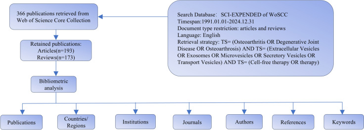
We obtained the study's bibliometric materials through the Science Citation Index Expanded (SCI-E) platform, accessed via Web of Science Core Collection (WoSCC) on January 18, 2025 [40]. To avoid bias, two investigators independently performed the literature search using the following search query: TS = (Osteoarthritis OR Degenerative Joint Disease OR Osteoarthrosis) AND TS = (Extracellular Vesicles OR Exosomes OR Microvesicles OR Secretory Vesicles OR Transport Vesicles) AND TS = (Cell-free therapy OR therapy). The search covered articles published from database inception to the search date. The study scope encompassed two types of publications: original research and review papers. From the WoSCC database, we obtained the full documentation and reference information for qualifying papers in plain text form. Key information including title, publication year, authors, journal, keywords, abstract, affiliations, and references was extracted for subsequent analyses. Any discrepancies in the search results were resolved through discussion and consensus.
Bibliometric analysis
The compilation of fundamental metrics—publication numbers, regional distribution, institutional data, journal details, and authorship information—was performed using Microsoft Excel 2021The journals' impact factors (IF) and disciplinary categories were extracted from the 2023 edition of Clarivate Analytics JCR rankings. The visualization and analysis of bibliometric data were accomplished through CiteSpace (version 6.4.R1) [3], VOSviewer (version 1.6.20) [4], and the R software "Bibliometric Package" (version 4.4.2). We also used Scimago Graphica to visualize the data.

Flowchart of literature screening in this study
Results
Analysis of publications

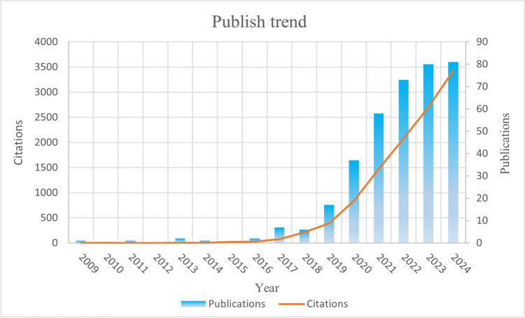
Annual publication volume and citation frequency of relevant articles
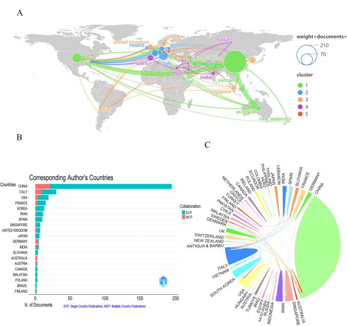
Contribution by countries/regions
Figure 3B indicates the collaboration patterns of corresponding authors by country, categorized as Single Country Publications (SCP, shown in cyan) and Multiple Country Publications (MCP, shown in red). The data shows that although major research countries including China, the United States and Italy maintain strong publication records, most of them are the results of domestic cooperation, and cross-border research partnerships are insufficient. In contrast, although Germany has a relatively small number of publications, it exhibits a positive trend in international cooperation.
Figure 3C illustrates the research collaboration network among different countries. Arc lengths represent publication volume, while line thickness indicates collaboration intensity, with thicker lines denoting more frequent collaboration. China occupies a substantial arc length, indicating significant research output, while multiple connecting lines converge at both China and the United States, identifying them as principal hubs of research collaboration.

Analysis of countries/regions.International collaboration network map. Node size represents publication volume, and countries with close collaborations are assigned the same color.Distribution of corresponding authors by country. Same-Country Publication (SCP), indicating the number of papers published by authors from the same country, serving as an indicator of domestic research collaboration. Multi-Country Publication (MCP), indicating the number of papers published by authors from different countries, representing an important measure of international research collaboration.Inter-country cooperation network. Sector area reflects research output, while line thickness indicates collaboration intensity A B C
| Rank | Countries/regions | Counts | Countries/regions | Citations | Countries/regions | total link strength |
|---|---|---|---|---|---|---|
| 1 | China | 202 | China | 6063 | USA | 41 |
| 2 | USA | 35 | Singapore | 1557 | China | 33 |
| 3 | Italy | 33 | USA | 1052 | Germany | 24 |
| 4 | France | 17 | France | 1031 | UK | 22 |
| 5 | South korea | 16 | Italy | 651 | Italy | 20 |
| 6 | Spain | 14 | South korea | 495 | Australia | 17 |
| 7 | Iran | 12 | Switzerland | 367 | Ireland | 16 |
| 8 | UK | 12 | Netherlands | 343 | Egypt | 14 |
| 9 | Australia | 10 | UK | 284 | France | 13 |
| 10 | Singapore | 10 | Australia | 245 | Switzerland | 13 |
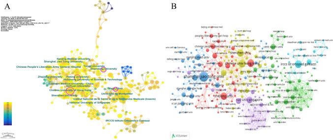
Contribution by institutions
Additionally, Fig. 4B presents the co-authorship analysis of institutions conducted using VOSviewer. Interestingly, two relatively independent clusters can be observed in the figure. The green cluster, centered around IRCCS Galeazzi Orthopedic Institute, has formed a tight European research network with IRCCS Rizzoli Orthopedic Institute, Université de Montpellier, and Université de Lorraine, demonstrating strong clinical research capabilities in orthopedics. The collaboration pattern of this cluster indicates a well-established clinical research framework. In contrast, the purple cluster, centered around the National University of Singapore, exhibits a unique academia-industry collaboration model, particularly through its connections with biotechnology companies such as Exocobio Inc and Zodiak Biosci Inc. The structure of this cluster reflects an emphasis on translational research and commercialization potential in extracellular vesicle therapy.

Analysis of institutional collaborations.CiteSpace network map. Nodes represent institutions with size indicating research output; lines show collaboration strength between institutions; purple rings highlight nodes with high betweenness centrality (BC ≥ 0.1), indicating key network intermediaries.VOSviewer co-authorship map. Node size reflects publication volume; connections indicate collaboration strength; institutions with close partnerships share the same color A B
Contribution by journals

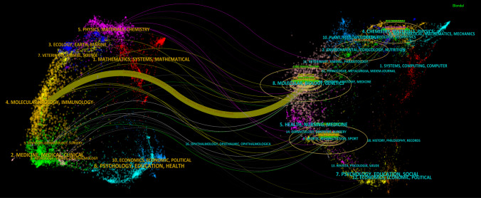
Analysis of academic journal citation networks using dual-map overlay. The left map shows citing journals, the right shows cited journals. Colored trajectories trace citation flows between research fields, with trajectory width indicating citation intensity—thicker paths represent stronger citation relationships
| Rank | Journal | Counts | IF (2023) | JCR | Cited-Journal | Citations | IF (2023) | JCR |
|---|---|---|---|---|---|---|---|---|
| 1 | International Journal of Molecular Sciences | 26 | 4.9 | Q1 | Biomaterials | 1,057 | 3.9 | Q2 |
| 2 | Frontiers in Bioengineering and Biotechnology | 19 | 4.3 | Q1 | Stem Cell Research & Therapy | 916 | 7.1 | Q1 |
| 3 | Stem Cell Research & Therapy | 12 | 7.1 | Q1 | Frontiers in Bioengineering and Biotechnology | 562 | 4.3 | Q1 |
| 4 | Frontiers in Cell and Developmental Biology | 11 | 4.6 | Q1 | Osteoarthritis and Cartilage | 492 | 7.2 | Q1 |
| 5 | Journal of Nanobiotechnology | 11 | 10.6 | Q1 | Scientific Reports | 458 | 3.8 | Q1 |
| 6 | Cells | 9 | 5.1 | Q2 | Theranostics | 451 | 12.4 | Q1 |
| 7 | Bioactive Materials | 8 | 18 | Q1 | International Journal of Molecular Sciences | 364 | 4.9 | Q1 |
| 8 | Journal of Orthopaedic Translation | 8 | 5.9 | Q1 | Seminars in Cell & Developmental Biology | 354 | 6.2 | Q1 |
| 9 | Biomedicines | 7 | 3.9 | Q2 | Journal of Nanobiotechnology | 350 | 10.6 | Q1 |
| 10 | Stem Cells International | 7 | 3.8 | Q2 | Bioactive Materials | 346 | 18 | Q1 |
Contribution by authors and co-cited authors

Analysis of author relationships.Co-authorship cluster map. Nodes represent authors with size indicating publication count; line thickness shows collaboration intensity; color-coded clusters represent closely collaborating research groups.Co-citation network. Node size reflects citation frequency or academic influence; line thickness indicates co-citation strength; same-colored authors are frequently cited together, suggesting similar research domains or complementary contributions A B
Analysis of keywords
Keywords are typically the most representative terms selected to explain research topics and serve as key indicators in bibliometrics for assessing research concentration and shifts in research focus. Analyzing keyword variations reveals the primary research directions and evolutionary patterns of research hotspots within a field. We extracted 744 keywords from 366 articles and used VOSviewer software to build a co-occurrence network, identifying 94 keywords that appeared at least three times. The keywords were automatically categorized into four main functional groups. Cluster 1 centers on the pathological mechanism research of osteoarthritis, encompassing related pathological processes such as cartilage damage, rheumatoid arthritis, and osteoporosis, reflecting the fundamental research foundation in this field; Cluster 2 focuses on extracellular vesicles and links to emerging therapeutic strategies including drug delivery, gene therapy, and cell-free therapy, demonstrating innovative developments in treatment approaches; Cluster 3 primarily revolves around mesenchymal stem cells and tissue engineering, closely connected with biomarker and senescence research, highlighting the significant role of stem cell therapy in the field of osteoarthritis; Cluster 4 concentrates on the clinical translation of regenerative medicine, including application research in biomaterials, platelet-rich plasma, and adipose-derived stem cells. Notably, extracellular vesicles, as an emerging therapeutic carrier, have formed close connections with all three other clusters. This multidirectional cross-correlation suggests the vast application potential of cell-free therapeutic strategies based on extracellular vesicles in future osteoarthritis treatment.
Figure 7B, constructed using CiteSpace software, illustrates the evolution trajectory and research hotspot distribution in this field over the past 15 years (2009–2024). The research themes demonstrate distinct temporal progression characteristics: early research primarily focused on articular cartilage pathology and basic research of apoptosis mechanisms; around 2015, there was a peak in cell therapy research represented by mesenchymal stem cells, while intra-articular injection drug delivery strategies and cartilage repair techniques gained widespread attention. Notably, after 2020, research related to extracellular vesicles rapidly emerged and formed close coupling with mesenchymal stem cell research, reflecting the transition from traditional cell therapy to cell-free therapeutic strategies. The knowledge map also reveals interactions among multiple research themes including synovial fluid microenvironment, cartilage defect repair, and angiogenesis, suggesting that osteoarthritis treatment research is moving towards a more comprehensive and precise direction. Particularly, as an emerging therapeutic carrier, extracellular vesicles display strong momentum in biogenesis mechanism research and delivery system development, which may indicate that cell-free therapy based on extracellular vesicles will play an important role in future osteoarthritis treatment. Author keywords typically represent the most representative terms chosen to explain research topics.

Keyword analysis.Trending topics. This visualization shows the temporal evolution of research themes based on keyword frequency changes over time.Timeline visualization of research trends. Keywords within the same cluster are arranged horizontally along the timeline, with earlier-appearing nodes representing foundational research areas, larger nodes indicating greater attention, and longer spans showing sustained research duration, thereby revealing the emergence, growth, and decline of research topics over time A B
Analysis of references

Reference analysis.Co-cited reference clustering. References with similar themes or research directions are grouped into clusters, each assigned distinct colors. The establishment and development of certain clusters may depend on literature from other clusters as their theoretical foundation or methodological source. These dependency pathways are visualized through arrows in the diagram, illustrating how knowledge flows from foundational works to subsequent research.Top 25 references with strongest citation burst. The blue bar represents the entire lifecycle of the keyword, while the red bar indicates the burst period of keyword research, detecting the start and end times as well as the burst intensity A B
| Rank | Title | Journal | First Author | Publication year | Total citations |
|---|---|---|---|---|---|
| 1 | Exosomes derived from human embryonic mesenchymal stem cells promote osteochondral regeneration | Osteoarthritis and Cartilage | Zhang, S | 2016 | 492 |
| 2 | Mesenchymal stem cells derived exosomes and microparticles protect cartilage and bone from degradation in osteoarthritis | Scientific Reports | Cosenza, Stella | 2017 | 446 |
| 3 | MSC exosomes alleviate temporomandibular joint osteoarthritis by attenuating inflammation and restoring matrix homeostasis | Biomaterials | Zhang, Shipin | 2019 | 376 |
| 4 | miR-100-5p-abundant exosomes derived from infrapatellar fat pad MSCs protect articular cartilage and ameliorate gait abnormalities via inhibition of mTOR in osteoarthritis | Biomaterials | Wu, Jiangyi | 2019 | 374 |
| 5 | MSC exosome as a cell-free MSC therapy for cartilage regeneration: Implications for osteoarthritis treatment | Seminars in Cell & Developmental Biology | Toh, Wei Seong | 2017 | 354 |
| 6 | Comparison of exosomes secreted by induced pluripotent stem cell-derived mesenchymal stem cells and synovial membrane-derived mesenchymal stem cells for the treatment of osteoarthritis | Stem Cell Research & Therapy | Zhu, Yu | 2017 | 308 |
| 7 | Mesenchymal Stromal/stem Cell-derived Extracellular Vesicles Promote Human Cartilage Regeneration ()In Vitro | Theranostics | Vonk, Lucienne A | 2018 | 267 |
| 8 | Exosomes from embryonic mesenchymal stem cells alleviate osteoarthritis through balancing synthesis and degradation of cartilage extracellular matrix | Stem Cell Research & Therapy | Wang, Yafei | 2017 | 244 |
| 9 | Chondrocyte-Targeted MicroRNA Delivery by Engineered Exosomes toward a Cell-Free Osteoarthritis Therapy | Acs Applied Materials & Interfaces | Liang, Yujie | 2020 | 239 |
| 10 | Exosome-mediated delivery of kartogenin for chondrogenesis of synovial fluid-derived mesenchymal stem cells and cartilage regeneration | Biomaterials | Xu, Xiao | 2021 | 230 |
Discussion
Basic information
Amidst the continual innovations in scientific scholarship, keeping pace with the latest research developments is more critical than ever. The proliferation of big data has led to an unprecedented volume of scholarly publications and data, resulting in growing difficulties for academics striving to monitor and assess recent scientific advancements. Nevertheless, by conducting statistical evaluations of scholarly information, bibliometric analysis offers a useful method for better understand research advancements, recognize growing areas of scientific interest, and forecast forthcoming scientific trajectories. From what we have gathered, this is the first bibliometric report on OA and EVs. Our analysis incorporated a sum of 366 scholarly articles, comprising 193 articles and 173 reviews. In terms of annual publication output, the period from 1991 to 2018 can be considered the first stage, characterized by a low number of publications, and no more than 10 papers were published in any given year. The years 2019 onwards mark the second phase, characterized by the steady worldwide emergence of top-tier studies in this domain. From 2022 onwards, the publication output has remained stable. The third stage began in 2022, when publication output stabilized and peaked in 2024. Among the main contributors in this area, China boasts the greatest quantity of scholarly output and total citations, closely followed by the United States, both being the principal drivers of research in this domain. Furthermore, it is noteworthy that notwithstanding the fewer publications, certain countries have an exceptionally elevated mean citations for each article. For instance, Singapore leads by a significant margin with an impressive 188.00, followed by Netherlands (165.00) and Ireland (76.00). The remarkably high average citation rate of Singapore can be ascribed to the issuance of various groundbreaking research reports by institutions such as the National University of Singapore and the Agency for Science, Technology and Research (A*STAR). For example, a study published in Biomaterials in 2018 found that exosomes extracted from MSCs improve cartilage repair through various mechanisms, including promoting proliferation, inhibiting apoptosis, and immunomodulation [33]. Toh et al. published a systematic review in Seminars in Cell & Developmental Biology in 2017, discussing the application prospects of MSCs-exos as cell-free therapeutics in cartilage regeneration and OA treatment [34]. In the same year, Wang et al. issued a scientific report in Stem Cell Research & Therapy, showing that exosomes originating from MSCs in embryos can mitigate OA by equalizing the production and breakdown of the extracellular matrix (ECM) of cartilage [46]. Additionally, Tan et al. published a systematic review in Tissue Engineering Part B in 2021 [47], summarizing the current status of MSCs-exos applications in animal studies of cartilage regeneration. From an institutional analysis perspective, Shanghai Jiao Tong University has published the highest number of papers. Despite this, it bears emphasizing that the extent of joint initiatives between academic establishments is comparatively minimal, with the majority of collaborations occurring at the country or territory level. In light of this, forging cooperative alliances among various academic organizations and investigative units is paramount for forthcoming studies. The periodical boasting the greatest quantity of printed articles is the International Journal of Molecular Sciences, while the journal with the highest number of citations is Biomaterials. De Girolamo, Laura from Aix-Marseille Universite and Ragni, Enrico from IRCCS Orthoped Inst Galeazzi have jointly published the highest number of papers, while Noel, Daniele from Universite de Montpellier had the highest citation rate.
Current research status and hot spots
Mechanisms of action of cell-free therapy based on EVs for OA
Cell-free therapy based on EVs has surfaced as a groundbreaking approach in regenerative medicine, leveraging the curative capacity of cell-secreted bioactive substances, as opposed to the cellular components themselves, to promote tissue restoration and regeneration [48, 49], and offering an alternative to traditional cell-based therapies that require direct transplantation of living cells into the target tissue [28]. These vesicles, including exosomes and MVs [50], made up of various functional substances, such as proteins, lipids, and nucleic acids [20], which transport to affected cells within injured joint structures, subsequently altering their responses and capabilities [51, 52]. Among these alternatives, vesicles released by MSCs have display favorable medicinal impacts in multiple test scenarios, such as heart-related illnesses, sudden kidney failure, hepatic impairment, and lung tissue damage [52 –56]. These vesicles influence immune system activity by decreasing the release of inflammation-related proteins, such as IL-1β and TNF-α, and by fostering an environment conducive to tissue regeneration [16]. Meanwhile, gene-modifying activities driven by miRNAs are essential for the medicinal properties of cell-free approaches, with EVs being enriched in miRNAs that can modulate the expression of genes involved in inflammation, autophagy, and other relevant pathways. Studies indicate these vesicles promote macrophage conversion from inflammatory M1 state to regenerative M2 form, supporting decreased joint inflammation [23, 57]. Additionally, studies reveal that EVs enhance cartilage matrix restoration in osteoarthritic tissue by reducing levels of proteins that break down matrix components, including MMP13 and ADAMTS5, thereby maintaining cartilage structure and delaying OA development [32, 46, 58, 59]. Moreover, EVs modify chondrocyte metabolic processes by boosting cellular energy production and ATP generation, thus strengthening their performance and stress resistance [60]. The cell homing properties of certain EVs, mediated by chemokines and other signaling molecules, allow for the recruitment of endogenous stem cells or progenitor cells to the site of injury, where they can participate in the tissue repair and regeneration process [61]. Some studies have also revealed that microparticles can upregulate the expression of type II collagen, suppress the expression of inflammatory markers and macrophage activation, protect chondrocytes from apoptosis, and improve cartilage parameters [30]. These microparticles exhibit therapeutic effects similar to those of exosomes, thus playing a positive role in the treatment of OA [62]. Beyond the direct actions of EVs, this treatment also enhances tissue regeneration through paracrine effects, with therapeutic cells secreting various forms of cytokines, growth factors, and other bioactive molecules that modulate the microenvironment of the damaged joint tissues, suppressing inflammation, promoting blood vessel formation, and modulating immune responses [63, 64]. Cell-free approaches target various OA mechanisms, offering an integrated solution for joint disease while advancing innovative regenerative treatments in orthopedic medicine [65, 66].
EVs in the treatment of osteoarthritis
OA, a common joint disorder, causes progressive degeneration that significantly diminishes patient well-being and daily functioning. As researchers seek to better understand the pathogenesis of this debilitating condition and develop more effective treatments, they have turned their attention to the role of EVs in the disease process. Exosomes and MVs, subclasses of EVs, have garnered significant attention in the pathogenesis and treatment of OA. The mechanisms underlying their roles have emerged as a focal point of research in this field. Cosenza et al. discovered that EVs derived from MSCs possess a chondroprotective effect, capable of mitigating IL-1β-induced chondrocyte apoptosis and matrix degradation [30]. Tao et al. identified key miRNAs, including miR-140-5p, EVs isolated from synovial fluid MSCs of individuals with OA. They found that these miRNAs promote chondrocyte proliferation and migration, suppress the expression of MMP-13 and ADAMTS-5, and are essential for EV-facilitated cartilage restoration [32]. Wu et al. further elucidated that MSC-derived EVs transport miR-100-5p and other miRNAs to inhibit the mTOR pathway in macrophages, thereby attenuating synovial inflammation and exerting an immunomodulatory mechanism [23]. Liu et al. further revealed that the lncRNA KLF3-AS1 in MSCs-exos could adsorb miR-206 by sponging, thus alleviating the suppressive impact of miR-206 on GIT1. Therefore, EVs mediate the restraining influence of the lncRNA-KLF3-AS1/miR-206/GIT1 axis [67]. Taken together, EVs, as essential intercellular communication mediators, regulate the functions of chondrocytes and immunomodulatory cells through the transfer of functional molecules such as non-coding RNAs. They are essential to the development and treatment of OA, emerging as a frontier and hotspot in precision medicine and regenerative medicine research.
Future directions
Exosome engineering
Exosomes, a type of vesicles secreted by cells, have developed into a novel drug delivery vehicle owing to their distinctive advantages in various aspects, such as biocompatibility, stability, and ability to cross biological barriers [68, 69]. Exosomes possess a multitude of unique properties, for example capability to cross biological boundaries, strong blood circulation persistence, and precise targeting ability, rendering them outstanding drug delivery vehicles. These distinctive features can be attributed to their biological composition and tiny dimensions, typically ranging from 40 to 100 nm [68, 70]. Engineered exosomes, which have undergone surface modifications including genetic engineering and chemical alterations, demonstrate improved targeting abilities. These adjustments elevate regional concentrations, reduced immunogenicity, minimal toxicity, and strong engineerability, thereby enhance their potential for treating numerous medical conditions [71]. Beyond serving as drug delivery vehicles, exosomes have natural properties that support osteoarthritic cartilage restoration. These inherent capabilities include the mitigation of local inflammation, the promotion of anabolic metabolism, and the suppression of catabolic processes [27, 72]. Several genes within chondrocytes have been identified as closely related to the progression mechanism of OA. Consequently, genome editing is considered a possible therapeutic approach that could offer long-term benefits for patients suffering from this debilitating condition [73, 74]. Liang et al. genetically engineered the exosomal surface protein Lamp2b to express a chondrocyte-affinity peptide on the exosome exterior, allowing vesicles to locate cartilage cells and be retained within the joint after intra-articular injection. Moreover, he combination of exosomes and lipid vesicles allowed the resulting hybrid exosomes to encapsulate CRISPR/Cas9 plasmids meanwhile maintaining the chondrocyte-homing properties, thereby delaying cartilage degradation in vivo and accelerating bone tissue repair [75].
Chen et al. researched the relationship between FGF18 silencing and the short of assorted cartilage-specific genes with in vivo and in vitro studies of OA progression. They s effectively created a innovative in vivo approach founded on CRISPR/Cas9 technology to activate the FGF18 gene in OA chondrocytes on a genetic basis. This method endowed chondrocytes with targeting capabilities, thereby enhancing in vivo binding efficiency and biosafety [76].
Zhao et al. utilized engineered exosomes derived from subcutaneous (SC) adipose tissue-derived MSCs-exos to achieve targeted transport of miR-199a-3p into chondrocytes. This approach was feasible due to the relative ease of obtaining healthy donor subcutaneous adipose tissue. In an OA mouse model, this targeted delivery system displayed considerable cartilage regeneration [77].
Wan et al. designed a novel engineered exosome-based drug delivery system to alleviate OA symptoms. They chemically modified exosomes by acylating methacrylic anhydride with the amino groups at the exosomal membrane, forming olefin double bond-modified exosomes. Through acylation with the peptides' amino groups, targeting peptides were conjugated to the modified exosomes, resulting in peptide-exosome binding. The engineered exosomes, modified with the cartilage-affinity peptide WYRGRL, were encapsulated in a new light-crosslinked spherical gelatin methacryloyl hydrogel (GelMA) for OA treatment. The performance of the engineered exosomes abundant in the small inhibitor LRRK2-IN-1 (W-Exo-L@GelMA) was surveyed both in cell cultures and animal models. W-Exo@GelMA evidenced effective chondrocyte targeting and markedly suppressed the expression of genes related to inflammation and immunity in OA [78]. These innovative, in-depth and extensive studies have laid an extremely solid foundation for the cell-free treatment of OA.
Looking ahead, several challenges remain. To advance exosome-centered OA therapy, standardization of exosome isolation and characterization protocols must be achieved, effective regulatory processes must be implemented, and therapeutic efficacy must be validated through clinical trials [79].
New drug delivery system for EVs
Recently, the development of novel delivery systems has become a focal point of investigation in OA therapy. The treatment approaches for OA can be divided into two primary groups: pharmacological and non-pharmacological interventions. Pharmacological therapies can be further subdivided based on the route of administration, which includes oral, injectable, and topical modalities [80]. An ideal drug delivery platform should efficiently cross biological barriers and precisely release the necessary quantity of active compounds at the designated time and location. Utilizing naturally derived materials with excellent biocompatibility for targeted drug delivery to specific cells or molecules ensures the safety of the drug delivery vehicle [81 –83].
The microenvironment of osteoarthritic joints is characterized by distinct pathological changes. These changes are relevant to factors such as pH levels, reactive oxygen species (ROS), and enzyme concentrations. Consequently, endogenous stimuli-responsive mechanisms that are sensitive to these conditions are currently under development. Nanoplatform-based drug delivery systems present innovative solutions for precise drug administration. These platforms enable enhanced drug accumulation and release kinetics at targeted disease locations, with the ability to respond to both endogenous and exogenous triggers for controlled therapeutic delivery. This strategy improves therapeutic uptake while reducing adverse effects throughout the body [81, 84]. To date, multiple delivery vehicles engineered for cartilage specificity have been created that facilitate the penetration of the compact ECM and improve therapeutic transport. Notably, nanoparticles incorporating chondrocyte-affinity peptides exhibit prolonged retention within the cartilage, causing them to become viable alternatives for the treatment of OA. In this burgeoning area of drug delivery study, the surface of exosomes is altered to boost their capabilities, facilitating site-specific drug delivery, along with in vivo visualization and tracing. Notably, nanoparticles incorporating chondrocyte-affinity peptides exhibit prolonged retention within the cartilage, making them promising candidates for the treatment of OA. Surface modification of exosomes in this innovative area of drug delivery research aims to augment their capabilities, facilitating targeted drug delivery, in vivo imaging, and monitoring [85].
Some researchers have attempted to establish an effective delivery strategy to enable exosomes to achieve continuous release in the superficial defect area [86]. According to a study carried out by Zhang et al., an injectable hydrogel with robust bonding properties to the moist chondral tissue was prepared through the enzymatic cross-linking of alginate-dopamine, chondroitin sulfate, and regenerated silk fibroin (AD/CS/RSF). The interfacial shear strength of the hydrogel reached 120 kPa. By encapsulating exosomes within the hydrogel, endogenous BMSCs in a rat model were recruited to the lesion location through chemoattractant transduction cascades and induced to differentiate into chondrocytes, assisting in the restoration of chondral lesions in the trochlear groove. This research has yielded an encouraging bioengineered substance for individuals with shallow chondral lesions, potentially deliverable via minimally invasive arthroscopic techniques [87]. Chen et al. designed a bioscaffold for the delivery of MSCs-exos and fabricated a 3D-printed scaffold composed of chondrocyte ECM, gelatin methacrylate (GelMA), and exosomes with radial channels utilizing desktop stereolithography technology. In a rabbit osteochondral lesion model, the ECM/GelMA/exosome scaffold productively rehabilitated chondrocyte mitochondrial dysfunction, increased chondrocyte motility, as well as polarized the response of synovial macrophages towards the M2 phenotype. The 3D printed scaffold remarkably promoted cartilage restoration in the in vivo study. The approach may serve as an encouraging approach for the therapy of OA [88]. Zeng et al. designed a multifunctional hydrogel system inspired by mussels, which exhibited thermosensitivity, self-healing properties, and adhesive characteristics. This system enabled the co-delivery and complementary actions of MSCs-exos and icariin (ICA). The intra-articular injection system combining ICA and MSCs-exos is expected to be retained within the joint cavity, alleviate cartilage atrophy, and ensure proper cartilage thickness [27].
Future research will focus on developing novel EVs-biomaterial composite carriers, optimizing 3D printing processes, and improving their loading capacity and bioavailability. Furthermore, EVs modification strategies, such as lipid insertion metabolic labeling, and peptide CAQK-modified, are important means to enhance the pharmacokinetic properties of EVs [89 –92]. Continued efforts to strengthen the fundamental research and industrialization exploration of exosome engineering, along with the establishment of standardized technical guidelines and translational pathways, will undoubtedly promote the widespread application of engineered exosomes in the precise diagnosis and treatment of OA. With the progress in nanotechnology, tissue engineering, and other fields, novel EVs delivery systems are anticipated to assume a more significant function in the identification and management of degenerative joint disease.
Conclusion
In conclusion, this article provided a thorough examination of the research outlook and trends in the domain of cell-free therapy based on EVs in OA. This bibliometric analysis also uncovers the key research themes and hotspots, and prominent the importance of interdisciplinary collaboration, spanning areas such as biomaterials, tissue engineering, and nanotechnology, in driving innovation and translational advances in cell-free for OA.
However, the study also identifies potential gaps and challenges, such as the optimization of EVs engineering techniques and the evaluation of long-term safety and efficacy in clinical settings. Future studies need to concentrate on dealing with these limitations and exploring novel strategies for harnessing the therapeutic potential of EVs in OA management.
In summary, this quantitative literature assessment offers an informative asset for investigators, healthcare professionals, and decision-makers by presenting an evidence-based outlook on the present landscape and forthcoming trajectories of cell-free therapy based on EVs in OA. The findings emphasize the importance of fostering international collaboration, investing in interdisciplinary research, and translating basic science discoveries into clinical applications. As the realm persists in evolving, it is expected that cell-free therapy based on EVs will exert an increasingly crucial role in the development of innovative, personalized, and efficacious therapies for OA, eventually enhance patient results and life quality.