
What this is
- This study investigates the cardioprotective effects of hydrogen gas (Hgas) inhalation in rat cardiomyocytes.
- It explores how Hgas activates cellular signaling pathways and to mitigate .
- The findings suggest that preconditioning with Hgas may be a viable preventive intervention for cardiac surgery patients.
Essence
- Preconditioning with inhaled Hgas protects rat cardiomyocytes from oxidative stress by activating the MAPK/ERK pathway and . This suggests potential clinical applications in preventing cardiac damage during surgery.
Key takeaways
- Hgas inhalation significantly inhibits increases in intracellular calcium concentration and mobilization from the endoplasmic reticulum, indicating a protective mechanism against oxidative stress.
- The expression of ERK and mitochondrial membrane potential levels were preserved with Hgas inhalation, while the inhibitor 3-methyladenine abolished these protective effects.
- The study demonstrates that Hgas inhalation activates and signaling pathways, providing a robust preconditioning effect against .
Caveats
- The study's findings are based on a high concentration of Hgas, and the effects of lower concentrations remain untested.
- The inhalation schedule may influence the residual effects of Hgas, as myocardial concentrations were not measured.
- The study primarily focused on exploratory research, necessitating further investigation into the dose-response relationship of Hgas in cardiac preconditioning.
Definitions
- ischemia/reperfusion injury: Tissue damage caused when blood supply returns to the tissue after a period of ischemia or lack of oxygen.
- autophagy: A cellular process that degrades and recycles cellular components to maintain homeostasis and respond to stress.
Simplified
Introduction
Ischemia/Reperfusion (I/R) injury is a major adverse event in patients undergoing cardiovascular surgery and is associated with massive production of reactive oxygen species (ROS) during reperfusion [1]. Most importantly, superoxide-derived hydroxyl radicals cause severe cellular damage because of their highly oxidizing properties. Hydrogen (H2) gas is a potential therapeutic agent against oxidative stress, because it selectively scavenges cytotoxic hydroxyl radicals but does not eliminate several ROS that possess physiological activities [2]. The feasibility and effectiveness of the inhalation approach could promote the clinical application of H2 gas in pathological states associated with massive ROS generation. Several clinical trials have mentioned that H2 gas inhalation can reduce I/R injury-induced myocardial damage in post-cardiac arrest syndrome or percutaneous coronary intervention [3, 4].
In addition to its scavenging properties against hydroxyl radicals, H2 gas can potentially activate several signaling pathways and mitigate I/R injury-induced myocardial damage [5]. Several signaling molecules, such as extracellular signal-regulated kinase (ERK) and Akt, are upstream factors that regulate the mitochondrial permeability transition pore (MPTP) to produce a preconditioning effect against subsequent cardiac damage [6, 7]. In addition to signaling pathways, autophagy has been recently shown to preserve mitochondrial homeostasis and confer cellular protection in cardiovascular diseases [8]. ERK and Akt are suggested to play major roles in regulating autophagy-related processes [9, 10]. Therefore, H2 gas inhalation may be a breakthrough treatment that induces the interaction of signaling pathways and autophagy to preserve cellular and mitochondrial homeostasis and confer the preconditioning effect in the myocardium.
Thus far, it is unclear whether H2 gas inhalation activates cellular cascades that mediate a preconditioning effect in the myocardium, although this intervention can potentially activate several signaling pathways. Similarly, it is controversial whether inhaled H2 gas activates autophagy-related processes. As very few researches have investigated the association between H2 gas and signaling cascades for modulating autophagy in the myocardium, this translational study aims to clarify whether preconditioning with inhaled H2 gas has a cardio-protective effect derived from the interaction between activated signaling pathways and autophagy. We hypothesized that H2 gas inhalation, together with the combined activation of cellular signaling cascades and autophagy, has a cardiac preconditioning effect against subsequent cardiac damage. Our findings could provide insight into the preconditioning effect of H2 gas against I/R injury-induced myocardial damage and the clinical significance of preventive interventions in perioperative medicine, especially in patients undergoing cardiac surgery.
Methods
Cell preparation
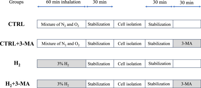
Six-week-old male Sprague-Dawley rats were purchased from an animal laboratory (CLEA Japan, Inc., Tokyo, Japan) and fed ad libitum under pathogen-free conditions until they attained a body weight of 300–350 g. In view of flammability, 3% concentration of H2 gas was prepared by mixing 4% H2 (Taiyo Nippon Sanso Co., Tokyo, Japan) and 100% oxygen (O2) in the ratio of 3:1. Prior to all experimental protocols, the rats inhaled 4 L·min−1 of 3% H2 or the mixture of nitrogen and O2 in the same ratio for 60 min in an experimental cage with an exhaust outlet, where H2 and O2 concentrations were continually monitored (GX-8000, Riken Keiki Co., Ltd., Tokyo, Japan).
After a 30-min stabilization, the hearts were harvested and isolated as previously described [11]. Briefly, rats were intraperitoneally administered with pentobarbital sodium (40 mg/kg, [Sigma-Aldrich, Inc., St. Louis, MO, USA]), xylazine hydrochloride (10 mg·kg−1, [Sigma-Aldrich, Inc.]), and heparin sodium (500 IU·kg−1, [Sigma-Aldrich, Inc.]). The isolated hearts were cannulated from the aorta and perfused with a normal Tyrode's solution consisting of 140 mM NaCl, 5.4 mM KCl, 1.8 mM CaCl2, 0.33 mM NaH2PO4, 0.5 mM MgCl2, 5.0 mM HEPES, and 5.5 mM glucose at 37 °C. After perfusion with a Ca2+-free Tyrode's solution containing 0.08% collagenase (FUJIFILM Wako Pure Chemical Co., Osaka, Japan), the left ventricle was minced and centrifuged at 400 ×g for 3 min. The pellet was repeatedly suffused and stabilized for 30 min at 23 °C (room temperature).
Experimental allocation

Time course of cell preparation and experimental allocation. CTRL: control; N: nitrogen; O: oxygen; MA: methyladenine; H: hydrogen 2 2 2
Fluorescence imaging for intracellular Caconcentrations 2+
To investigate increase in the intracellular Ca2+ concentration in response to oxidative stress, a major cause of I/R injury [12], fluorescence imaging with Fluo-4 acetoxymethyl ester (Dojindo Laboratories, Kumamoto, Japan), a specific calcium indicator, was conducted using a confocal laser microscope (LSM700; Carl Zeiss, Jena, Germany). Cardiomyocytes were incubated with 5 μM Fluo-4 acetoxymethyl ester for 30 min and then perfused with 2 ml·min−1 of the normal Tyrode's solution containing 100 μM of hydrogen peroxide (H2O2; [FUJIFILM Wako Pure Chemical Co.]) in the recording chamber at 23 °C. This concentration of H2O2 was shown to feasibly produce calcium overload in cardiomyocytes [13]. Viable rod-shaped cells were selected through a 40× objective lens and intermittently illuminated at a wavelength of 488 nm for 10 min. Time-course recordings were obtained from emissions above 515 nm to estimate the increasing intensity of Fluo-4 fluorescence throughout the illumination.
To investigate H2-induced modification of calcium kinetics, Ca2+ mobilization from the endoplasmic reticulum (ER) was estimated using fluorescence imaging [14]. Fluo-4-loaded cardiomyocytes were perfused with the Ca2+-free Tyrode's solution for 5 min at 23 °C, followed by the solution containing 10 mM caffeine (Sigma-Aldrich, Inc.) for 1 min. ER-stored Ca2+ is released through ryanodine receptors and increases the intracellular Ca2+ transient. Time-course recordings were obtained throughout the perfusion, with the peak value estimated as the ER-stored Ca2+ content. Furthermore, the decay of the Ca2+ transient was estimated using a time constant (τ) fitted to a single exponential, considered as an excretion function of intracellular Ca2+ through the Na+-Ca2+ exchange system [15].
Determination of MPTP opening
To investigate H2-induced modification of MPTP opening, fluorescence imaging was performed using tetramethylrhodamine ethyl ester (TMRE; [FUJIFILM Wako Pure Chemical Co.]) [11]. After incubation with 2 μM TMRE for 15 min, cells were perfused with the normal Tyrode's solution and intermittently illuminated at a wavelength of 543 nm for 10 min at 23 °C. Time-course recordings were obtained from emissions above 570 nm through a 40× objective lens to estimate the time of MPTP opening. The opening was determined by a sharp increase in TMRE fluorescence, which is effluent from the mitochondria and is dequenched [12].
Flow cytometry assays for signaling molecules and autophagy
To determine the upstream pathways relevant to H2-induced preconditioning effects, we examined the expressions of ERK and Akt, which are crucial kinases for promoting survival cascades [6, 7]. After exposure to the Ca2+-free or normal (1.8 mM Ca2+) Tyrode's solution for 30 min, the cells were fixed and incubated with an Alexa Flour 488®-labeled specific antibody for ERK1/2 or Akt (Phosphoflow; Becton Dickinson Biosciences, Bedford, MA, USA) according to the manufacturer's instructions. These antibodies are known to bind exclusively to phosphorylated residues, which are active forms of these molecules. The loaded cells were excited at a wavelength of 488 nm, filtered at 511 nm using flow cytometry (FACS II; Becton Dickinson, Bedford, MA, USA), and sorted on the basis of the forward and side scatter [16]. The expression of ERK or Akt was determined from the mean intensity of each fluorescence signal under a cellular distribution.
The emission from a low concentration of TMRE is well correlated with mitochondrial membrane potentials (ψm) [16]. After exposure to the Ca2+-free or normal Tyrode's solution, cardiomyocytes were incubated in the solution containing 100 nM TMRE and were then excited at a wavelength of 488 nm and were filtered at 511 nm using flow cytometry. ψm was estimated using the mean intensity of TMRE fluorescence under a cellular distribution.
To clarify H2-induced activation of autophagy, we examined the expressions of autophagosomes and microtubule-associated light chain (LC) 3-II, an integral factor activating autophagy-related processes [17, 18]. Cells were exposed to the Ca2+-free or normal Tyrode's solution and were then incubated with 1 μM DALGreen (Dojindo Laboratories), an indicator of autophagosome formation [19] or a Fluorescein-Isothiocyanate-labeled LC3-II antibody (Funakoshi Co., Ltd., Tokyo, Japan). The expressions of DALGreen and LC3-II were determined using flow cytometry as described above.
Statistical analysis
All analyses were conducting on the basis of per protocols and cells which did not complete the appropriate protocols were excluded from the data analysis. The sample size was determined based on previous findings in each experimental protocol to provide a power of α error of 0.05 and β error of 0.1. In the analyses of intracellular Ca2+ levels, a sample size of 15 was sufficient to detect a mean difference (MD) of 40% and standard deviation (SD) of 25% [13], whereas, for the opening of the MPTP, the number of 30 was sufficient to detect the difference in survival rates between 0.25 and 0.7 in the experimental period [11]. In flow cytometry analyses, the sample size of 8 was needed for the expression of signaling molecules, with an MD of 23% and SD of 10% [9]. All experimental protocols were not adopted to repetitive measurements from the view of experimental characteristics. Experimental data were transformed into rates of change from baseline values except for those of MPTP opening. After verifying normality and homoscedasticity, between-group comparisons were conducted using a one-way analysis of variance (ANOVA) and post-hoc Tukey's test. Mixed-effects models were used to conduct between-group comparisons, with the subject number as a random effect and the increasing rate at each point and experimental groups as a fixed effect. In contrast, repeated-measures ANOVA and post-hoc Holm's test were performed for within-group comparisons. The time to MPTP opening was assessed using Kaplan–Meier's curves and the log-rank test. Results are appropriately expressed as means ± SD, whereas those from the log-rank test are presented as medians [95% confidence intervals]. The number of experiments and rats are expressed as n and N. All statistical analyses were performed using the R software (version 4.0.3; R Foundation, Vienna, Austria), and data with a P-value less than 0.01 (two-tailed) were considered to be statistically significant in consideration of multiple comparisons.
Results
Changes in intracellular Caconcentrations 2+

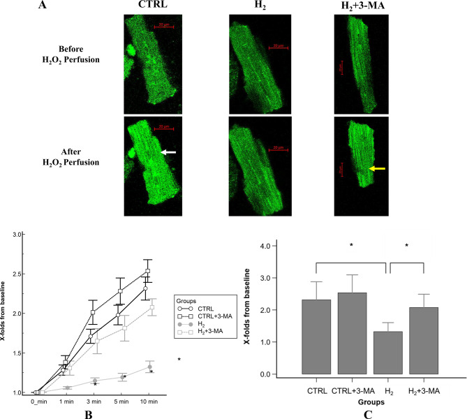
Changes in intracellular Calevels with 10-min HOperfusion. () Representative images of fluo-4-loaded cardiomyocytes before and after 10-min HOperfusion. A white arrow indicates that a cell emits intense luminescence. A yellow arrow indicates a cell produces moderate luminescence and cell-shrinking. () Time-course changes in the mean Fluo-4 fluorescence intensity during HOperfusion. Error bars show standard error of the mean. The mixed-effect models revealed statistical significance between the CTRL and Hgroups. () Comparison of the Fluo-4 fluorescence intensity after 10-min HOperfusion. Bar graphs show the increasing rate of Fluo-4 fluorescence from baseline. The rate in the Hgroup significantly decreased relative to those in the CTRL and H + 3-MA groups. *: statistical significance with < 0.001 ( = 15, = 5 for each experimental group). CTRL: control; H: hydrogen; MA: methyladenine; HO: hydrogen peroxide; n: the number of experiments; N: the number of rats 2+ 2 2 2 2 2 2 2 2 2 2 2 2 2 2 A B C p n N

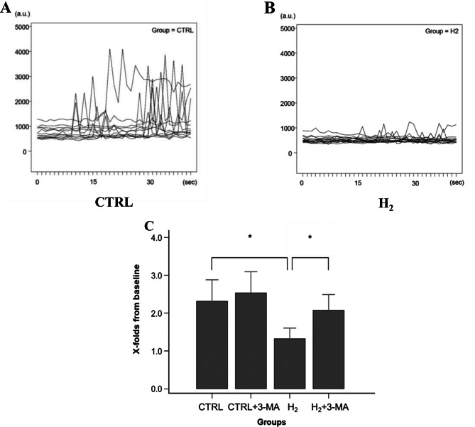
Estimation of intracellular Calevels after caffeine administration. (,) Time-course profiles of fluo-4 fluorescence after caffeine administration. () Comparison of Catransients from the ER. Bar graphs show the peak ratio of fluo-4 fluorescence from baseline. The ratio in the Hgroup significantly decreased relative to those in the CTRL and H + 3-MA groups. *: statistical significance with < 0.001 ( = 15, = 5 for each experimental group). CTRL: control; H: hydrogen; MA: methyladenine; ER: endoplasmic reticulum; n: the number of experiments; N: the number of rats 2+ 2+ A B C 2 2 2 p n N
Time analysis of MPTP opening

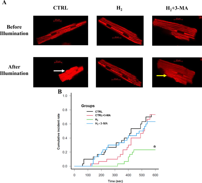
Time analysis of TMRE fluorescence for the opening of MPTP. () Representative images of the TMRE-loaded cardiomyocytes before and after 10-min perfusion. A white arrow indicates a cell produces intense luminescence and cellular hypercontraction. A yellow allow indicates that a cell produces moderate luminescence and cell-shrinking. () Kaplan–Meier curves of the opening of MPTP. The log-rank test revealed that the Hgroup had a significantly longer time to MPTP opening relative to the CTRL and H + 3-MA groups. *: statistical significance with < 0.01 for the CTRL and H + 3-MA groups ( = 30, = 10 for each experimental group). CTRL: control; H: hydrogen; MA: methyladenine; TMRE: tetramethylrhodamine ethyl ester; MPTP: mitochondrial permeability transition pore methyladenine; n: the number of experiments; N: the number of rats A B 2 2 2 2 p n N
Preserved signaling molecules and ψm levels
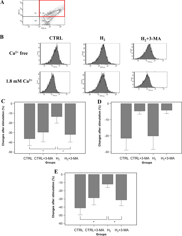
Regarding changes in the levels of ψm, Ca2+ stimulation remarkably decreased that of the CTRL group, whereas it did not affect that of the H2 group (CTRL, −41.1 ± 8.5%; H2, −11.9 ± 4.2% vs. Ca2+-free exposure; p < 0.001; Fig. 5E). The addition of 3-MA significantly decreased the ψm level relative to that of the H2 group (H2 + 3-MA, −31.3 ± 7.1%; vs. Ca2+-free exposure; p < 0.001 for H2: Fig. 5E).

Determination of activated signaling molecules and preserved mitochondrial electrical potentials. () A representative image of cell sorting using flow cytometry. A red square shows the studied filed. () Representative profiles of cellular distribution based on ERK expression with or without 1.8 mM Castimulation. () Changes in ERK expression after Castimulation. Bar graphs represent percentage changes in the mean values of ERK expression. The Hgroup preserved ERK expression, whereas the CTRL and H + 3-MA groups showed remarkably reduced ERK expression. *: statistical significance with < 0.001 ( = 8, N = 4 for each experimental group) () Changes in AKT expression after Castimulation. Bar graphs represent percent changes in the mean values of AKT expression. The Hgroup showed remarkably reduced AKT expression, which was similar to the CTRL group. The addition of 3-MA did not affect AKT expression irrespective of Hinhalation. () Changes in ψm levels after Castimulation. Bar graphs represent percent changes in the mean values of ψm. The Hgroup relatively preserved the level of ψm, whereas the CTRL and H + 3-MA groups remarkably decreased the ψm levels*: statistical significance with < 0.001 ( = 8, = 4 for each experimental group). CTRL: control; H: hydrogen; MA: methyladenine; ERK: extracellular signal-regulated kinase; ψm: mitochondrial membrane potentials; n: the number of experiments; N: the number of rats A B C D E 2+ 2+ 2+ 2+ 2 2 2 2 2 2 2 p n p n N
Increased autophagosomes and LC3-II expression

Determination of autophagy and autophagosome activation. () Representative profiles of cellular distribution based on DALGreen expression. () Changes in autophagosome formation after Castimulation. Bar graphs represent percent changes in the mean values of DALGreen. DALGreen expression was significantly increased in the Hgroup relative to the CTRL and H + 3-MA groups. *: statistical significance with < 0.001 ( = 8, = 4 for each experimental group) () Changes in LC3-II expression after Castimulation. Bar graphs represent percent changes in the mean values of LC3-II expression. LC3-II expression was significantly increased in the Hgroup relative to the CTRL and H + 3-MA groups. *: statistical significance with < 0.001. ( = 8, = 4 for each experimental group). CTRL: control; H: hydrogen; MA: methyladenine; LC: light chain; n: the number of experiments; N: the number of rats A B C 2+ 2+ 2 2 2 2 2 p n N p n N
Discussion
The current study demonstrated that preconditioning with 3% H2 gas for 60 min effectively inhibited the H2O2-perfused calcium overload and Ca2+ mobilization from the ER and prolonged to the time to MPTP opening. Furthermore, H2 gas inhalation preserved the expression of upstream signaling molecules of ERK and LC3-II and the electrical potentials in the mitochondria. These protective effects were completely abolished by pretreatment with the autophagy inhibitor 3-MA. These findings are congruent with our hypothesis that H2 gas inhalation has a cardiac preconditioning effect with the combined activation of cellular signaling cascades and autophagy and are novel in that H2 gas possesses a cellular mechanism for alleviating oxidative stress other than scavenging hydroxyl radicals.
Following the demonstration that H2 gas was a selective radical scavenger for hydroxyl radicals and alleviated oxidative stress during I/R injury [2], several clinical trials have shown that H2 gas inhalation is a successful treatment for I/R injury-induced cardiac damage [3, 4]. As previous studies mainly focused on concurrent or subsequent administration of H2 gas for ischemic events, preventive intervention with H2 gas inhalation–a more practical approach–remains unconfirmed owing to limited knowledge. This study demonstrated that prior inhalation of 3% H2 gas had a preconditioning effect against oxidative stress-induced myocardial damage and was mainly attributed to suppressing the increase in intracellular Ca2+ levels and Ca2+ mobilization from the ER. This action mechanism differs from scavenging hydroxyl radicals and leads to preservation of ψm and resistance to MPTP opening, which is strictly regulated by intracellular Ca2+ levels and the upstream signaling molecules [6, 7]. Taken together, the root mechanism of the preconditioning effect induced by H2 gas is to preserve mitochondrial homeostasis through the maintenance of intracellular Ca2+ levels and activate cellular survival pathways, including ERK. These findings were consistent with the previous report that some physiologically-active gas exerted a cardio-protective effect through the activation of ERK [20].
Several studies have mentioned the relationship between autophagy and cardio-protection, with which several signaling pathways are involved. The mitogen-activated protein kinase (MAPK)/ERK pathway promotes autophagy to remove waste products and maintain intracellular homeostasis [9]. In contrast, the phosphatidylinositol-3 kinase (PI3K)/Akt pathway inhibits autophagy to avoid excessive degradation of organelles [10, 21]. Despite their discrepant involvement with the autophagy process, both signaling pathways are key factors in cardiac preconditioning [6, 7]. On the basis of the significant increase in autophagosome formation and LC3-II expression, H2 gas inhalation predominantly induced autophagy with ERK activation in this study. However, positive Akt expression, a counterpart of ERK activation, was not induced by H2 gas inhalation. This might be attributable to our experimental feature of cardiac preconditioning, because H2 gas inhalation was completed before myocardial damage occurred. In fact, H2 gas inhalation can activate the PI3K/Akt pathway to inhibit autophagy, provided that cellular damage preceded the inhalation [22].
The current study demonstrates the preconditioning effect of H2 gas inhalation mediated through the combined effects of the MAPK/ERK pathway and autophagy. Besides ERK, autophagy can regulate MPTP opening by preserving mitochondrial homeostasis associated with potassium channels [23]. ERK is considered as an upstream factor in the development of autophagy [6]; however, we demonstrated the mutual interaction between ERK and autophagy because an autophagy inhibitor completely suppressed H2-induced ERK expression. Thus, H2 gas inhalation concurrently activates both systems and profoundly preserves cellular homeostasis, thereby producing a robust preconditioning effect in the myocardium. Recent studies suggesting the interaction between the MAPK/ERK pathway and autophagy support our findings and indicate a potential treatment target for several pathological conditions [10, 24].
The current study has some limitations. First, a 30-min stabilization duration after H2 gas inhalation was determined for the memory phase of cardiac preconditioning that is needed for the development of cellular signaling pathways [25]. The residual effect as a radical scavenger may have been related to the inhalation schedule because the myocardial concentrations of H2 were not measured. However, as H2 gas reportedly disappears from the myocardium minutes after discontinuing inhalation, it could not serve as a radical scavenger in this study [26]. Second, lower concentrations of H2 gas may have produced similar results but were not investigated in this study. A concentration of 3% was determined for safety margin and carrier gas use, although the dosage is generally restricted to 4% for inhalational use [27]. We primarily focused on exploratory research to assess the effectiveness of inhaled H2 gas at a relatively high concentration; therefore, further research on the dose-response effects of inhaled H2 gas on cardiac preconditioning is warranted. Third, the stored content of Ca2+ in the ER is known as the mainstream of calcium kinetics at I/R injury, heavily released into the cytosol at reperfusion [28, 29]. However, the influx of extracellular Ca2+ through the reverse-mode Na+-Ca2+ exchange (NCX) or transient receptor potential canonical channels might have been involved in the increased intracellular Ca2+ concentration in this study [13]. Furthermore, the conditions of the reverse-mode NCX might influence our findings that H2 gas inhalation did not change the excretion of intracellular Ca2+ through the forward-mode NCX. Fourth, we assessed the expression of ERK and Akt as representative molecules that generate cellular survival cascades in cardiomyocytes. The Janus kinase/signal transducer and activator of transcription pathway reportedly possesses a late-phase preconditioning effect after cytokine stimulation [30]. This finding contradicts our premise that the early preconditioning effect occurs within several hours after H2 gas inhalation. Fifth, western blot analysis is a gold standard of estimating signaling molecules, but it includes outcomes from dead cells induced by stimulation and may underestimate those form our experimental protocols using isolated cells [31, 32]. We therefore conducted a sequence of flow cytometry analyses and obtained results from the sorted cardiomyocytes with living potential.
Conclusions
The preconditioning with inhaled H2 gas confers a cardio-protective effect against oxidative stress, mediated through the activation of the MAPK/ERK pathway, but not the PI3K/Akt, pathway. Autophagy activation, which helps maintain cellular or mitochondrial homeostasis, facilitates the expression of the H2-induced survival cascades. Preventive intervention with H2 gas is valuable for ensuring feasibility and safety and has clinical significance in perioperative medicine, especially in patients undergoing cardiac surgery.