
What this is
- This research investigates the relationship between , α-Klotho protein levels, and cognitive performance in Alzheimer's disease (AD).
- The study includes 296 participants, categorized into groups based on cognitive status: AD dementia, amnestic mild cognitive impairment (), and cognitively unimpaired controls.
- Findings suggest that may be linked to lower odds of and AD dementia, particularly affecting memory performance in patients.
Essence
- is associated with lower odds of being classified as or dementia due to AD, particularly enhancing memory performance in patients carrying the APOE ε4 allele.
Key takeaways
- carriers showed a 61% lower likelihood of being classified as due to AD compared to cognitively unimpaired individuals, suggesting a potential protective effect.
- Among patients, KLOTHO-VS heterozygotes displayed better memory performance, especially in those also carrying the APOE ε4 allele, indicating a possible buffering effect against cognitive decline.
- No significant differences in α-Klotho protein levels were found between study groups, suggesting that while may influence cognitive outcomes, soluble α-Klotho levels did not correlate with memory performance.
Caveats
- The cross-sectional design limits causal inferences, making it difficult to determine the direction of associations between KLOTHO-VS and cognitive performance.
- The sample size was relatively small, leading to limited statistical power and potentially affecting the generalizability of the findings.
- Post-hoc power estimates indicated a bias towards Type II error, meaning null findings may reflect insufficient power rather than a true lack of association.
Definitions
- KLOTHO-VS heterozygosity: A genetic variant of the KLOTHO gene associated with various neuroprotective effects, particularly in Alzheimer's disease.
- α-Klotho: A protein encoded by the KLOTHO gene, involved in regulating aging and cognitive functions.
- amnesic mild cognitive impairment (aMCI): A condition characterized by noticeable memory problems that are greater than expected for a person's age but not severe enough to interfere significantly with daily life.
Simplified
Background
α-Klotho, encoded by the KLOTHO gene, is a single-pass transmembrane protein primarily expressed in the brain's choroid plexus and the kidney's distal tubule cells [1]. A soluble form of α-Klotho (sαKl) is produced through proteolytic cleavage from membrane-bound α-Klotho and released into the bloodstream or cerebrospinal fluid (CSF), where it may act as a hormone exerting multiple systemic biological actions on cells or tissues that do not express α-Klotho [2].
α-Klotho's role in the aging process was first identified through a study on KLOTHO knockout mice, whereby poor KLOTHO gene expression was shown to significantly reduce life expectancy through mechanisms similar to those seen in human aging [3]. Later research focusing on the central nervous system of the transgenic mice revealed that they also exhibited age-related neurodegenerative changes, including synaptic loss, neuronal degeneration, and cognitive impairment [4, 5]. On the other hand, overexpression of α-Klotho in mice was found to extend lifespan, improve learning, memory, synaptic plasticity and reduce amyloid-β (Aβ) burden [6 –9]. While transgenic mice models provided compelling evidence for α-Klotho's neuroprotective potential, human studies offer a crucial bridge to understanding its in vivo relevance in neurodegenerative diseases.
KLOTHO-VS heterozygosity (KL-VSHET), a functional genotype of the KLOTHO gene, has been associated with various beneficial effects, particularly in the context of AD and cognitive function [6, 10 –16]. Specifically, having the KL-VSHET haplotype has been shown to significantly decrease the risk of AD, although the effect appears to be restricted to carriers of the Apolipoprotein E ε4 allele (APOE ε4) [11]. This finding may be a function of the ability of the KL-VSHET to attenuate the adverse effects of APOE ε4 on Aβ accumulation and tau burden [10 –12, 14, 15, 17]. Additionally, evidence suggests that KL-VSHET is associated with better memory even in Aβ driven pathology, likely due to lowering the levels of tau protein [14]. One of the primary mechanisms by which KL-VSHET may confer resilience against AD is by increasing the levels of both CSF and serum sαKl [6, 18, 19].
While the promising associations between higher sαKl levels and better cognitive performance are evident in normal aging [20, 21], its role in AD remains unclear. Studies in AD patients link lower CSF sαKl to worse AD pathology and cognitive function [18, 22, 23]. In contrast, plasma sαKl shows inconsistent results, with some studies finding no difference between AD patients and controls [18, 24] and others reporting higher levels in AD patients [25]. This study extends prior work by assessing domain-specific cognition in biomarker-confirmed AD groups and evaluating both sαKl protein levels and KL-VS haplotype.
Based on previous research, we investigated whether KL-VSHET would be associated with a lower likelihood of aMCI or dementia due to AD compared to controls in APOE ε4 carriers vs. non-carriers. We expected positive results in APOE ε4 carriers but not in APOE ε4 non-carriers. We also tested whether KL-VSHET carriers would have better cognitive performance than those who were KL-VS non-carriers (KL-VSNC). Given prior evidence suggesting that KL-VSHET mitigates AD pathology predominantly in APOE ε4 carriers, we hypothesized that associations between KL-VSHET and both cognitive performance and likelihood of classification as AD would be stronger in individuals carrying the APOE ε4 allele. This expectation was grounded in multiple large-scale studies demonstrating that KL-VSHET attenuates APOE ε4–associated Aβ accumulation and cognitive decline in cognitively normal individuals at genetic risk for AD. These protective associations appear to be APOE ε4-dependent, with most studies reporting little to no benefit of KL-VSHET in APOE ε4 non-carriers [10, 12, 17]. Our study sought to extend this line of research by evaluating whether such APOE ε4-specific effects would persist across clinically defined stages of AD. Additionally, we sought to compare sαKl concentrations in CSF and in serum across patients with AD dementia, aMCI due to AD, and cognitively unimpaired individuals. We hypothesized that patients with AD dementia and with aMCI due to AD would exhibit lower levels of sαKl in CSF compared to cognitively unimpaired controls. Finally, we investigated whether CSF and serum sαKl would be associated with better cognitive performance.
Methods
Participants
Participants were drawn from the Czech Brain Aging Study cohort [26] at the Memory Clinic of Charles University/Motol University Hospital, and the Department of Neurology, Motol University Hospital in Prague, Czech Republic. All Czech Brain Aging Study participants are subjected to clinical and laboratory evaluations within three months of the initial visit, including routine blood tests, comprehensive neuropsychological assessment, and brain magnetic resonance imaging (MRI; 1.5 or 3 T with MP RAGE sequences). All participants involved in this study signed written informed consent approved by the Motol University Hospital ethics committee. All participants included in the study were White and of Czech nationality.
Inclusion/exclusion criteria
To be included in the present study, participants were required to meet criteria for one of three diagnostic groups—AD dementia, aMCI due to AD, or cognitively unimpaired controls—and to have either sαKl protein levels measured in CSF and/or serum or genotyping data available for the KL-VS haplotype. Participants were excluded if they had pre-existing neurological or psychiatric conditions that could impair cognitive function, including Parkinson disease, Lewy body dementia, frontotemporal lobar degeneration, psychosis, substance abuse, depression (≥ 6 points on the 15-item Geriatric Depression Scale) [27], stroke, traumatic brain injury, or multiple sclerosis. Additionally, patients with severe white matter vascular lesions on MRI (Fazekas score > 2 points) [28] were excluded. Participants suffering from renal failure (defined as a GFR min/1.73 m² or chronic kidney damage) were also excluded since kidneys are a major site of α-Klotho production. Participants were also excluded from the study sample due to KL-VS homozygosity, as this genotype is rare in the general population and would not permit meaningful statistical analysis.
Study sample
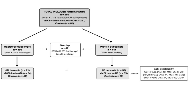
An overview of the composition of the two subsamples used in this study is shown in Fig. 1. Following application of these inclusion and exclusion criteria, a total of 296 participants had relevant data available and were included in the present study. Of the 296 participants, 231 were referred to the Memory Clinic by general practitioners, neurologists, or geriatricians based on cognitive difficulties reported by themselves or their informants. These participants comprise the diagnostic groups of AD dementia and aMCI due to AD used in both the haplotype and protein subsamples (see Fig. 1). The remaining 65 participants were cognitively unimpaired controls.
Based on available genetic and biomarker data, participants were divided into two partially overlapping analytic subsamples: one with available KL-VS haplotype (haplotype subsample), and one with available sαKl protein measurements in CSF and/or serum (protein subsample).
The haplotype subsample consisted of 196 participants and was used to examine the association between KL-VSHET and odds of AD as well as cognitive performance. It included 71 participants with AD dementia [29], 84 with aMCI due to AD [30], and 41 cognitively unimpaired controls. AD biomarker status in this subsample was determined using CSF (n = 129), Aβ PET imaging (n = 96), or both modalities (n = 29).
The protein subsample consisted of 147 participants and was used to investigate differences in sαKl levels across clinical groups and their associations with cognitive outcomes. It comprised 58 participants with AD dementia [29], 59 with aMCI due to AD [30], and 30 cognitively unimpaired controls. CSF sαKl levels were available for 131 participants (AD dementia n = 48, aMCI due to AD n = 55, controls n = 28), and serum sαKl levels were available for 118 participants (AD dementia n = 44, aMCI due to AD n = 46, controls n = 28). Both CSF and serum sαKl measurements were available for 102 participants (AD dementia n = 34, aMCI due to AD n = 43, controls n = 25). AD biomarker status in this subsample was determined using CSF (n = 147), Aβ PET imaging (n = 42), or both modalities (n = 42).
There was partial overlap between the two subsamples, with 47 participants contributing data to both analyses (AD dementia n = 21, aMCI due to AD n = 18, controls n = 8).

Flow diagram illustrating the composition of the study sample. A total of 296 participants were included based on the availability of eitherhaplotype genotyping or sαKl protein data. Two partially overlapping subsamples were derived: the Haplotype Subsample ( = 196), consisting of participants withhaplotype data, and the Protein Subsample ( = 147), consisting of participants with CSF and/or serum sαKl protein concentrations. A total of 47 participants were included in both subsamples. Each subsample was categorized by diagnostic group: AD dementia, aMCI due to AD, and cognitively unimpaired controls. Diagnostic group assignment required biomarker confirmation. All participants with AD dementia or aMCI due to AD were CSF and/or PET positive; all cognitively unimpaired controls were CSF and/or PET negative KL-VS n KL-VS n
Diagnostic information
Participants diagnosed with AD dementia met the NIA-AA 2011 criteria for dementia due to AD [29] based on progressive decline in at least two cognitive domains (i.e. ≥1.5 standard deviations (SD) lower memory test score than the age- and education-adjusted norms as well as similarly low score in at least one non-memory cognitive test), significant impairment in the ability to perform daily activities, and biomarker evidence of AD pathophysiology. Biomarker confirmation was established via either lumbar puncture or Aβ positron emission tomography (PET) imaging using 18 F-flutemetamol. Positive biomarker status was defined as CSF Aβ42 < 620 pg/mL and phosphorylated tau at amino acid 181 > 61 pg/mL and/or a positive Aβ PET scan.
Participants diagnosed with aMCI due to AD met the NIA-AA 2011 criteria for MCI [30] based on subjective reports of memory decline compared to their prior level, objective evidence of memory impairment (i.e. ≥1.5 SDs lower score than the age- and education-adjusted norms in any memory test), preserved independence in daily activities, and the absence of dementia. As with the AD dementia group, aMCI participants were also required to have biomarker confirmation of AD pathology, based on either CSF analysis (Aβ42 < 620 pg/mL and phosphorylated tau > 61 pg/mL) or a positive Aβ PET scan.
Cognitively unimpaired individuals were required to exhibit normal cognitive function (scores > 1.5 SD above age- and education-adjusted norms on all cognitive tests) and normal AD biomarkers in CSF and/or PET. These participants were recruited from two sources:
among individuals initially referred to the Department of Neurology, Motol University Hospital, for lumbar puncture to rule out central nervous system inflammation. After normal CSF analysis, absence of systemic inflammation, and no evidence of hippocampal atrophy on MRI, these individuals were enrolled into the Czech Brain Aging Study as cognitively unimpaired controls.among Czech Brain Aging Study patients followed for subjective cognitive decline. Although they reported cognitive difficulties that led them to seek medical evaluation, they showed unimpaired activities of daily living and normal cognitive function [31]. MRI findings revealed no hippocampal atrophy, and AD biomarkers in CSF or PET were within normal ranges.
Genotyping
APOE and KLOTHO genotypes were determined at the Department of Clinical Biochemistry, Hematology and Immunology, Homolka Hospital, Prague, Czech Republic.
DNA isolation was performed by Zybio Nucleic extraction kit WB-B from whole blood samples according to manufacturer's protocol (Zybio, Chongqing, China).
APOE genotyping was performed according to IdahoTech protocol (Luna Probes Genotyping Apolipoprotein [ApoE] Multiplexed Assay) for high-resolution melting analysis (HRM) [32, 33].
The analysis of KL-VS haplotype was done also by HRM analysis of single nucleotide polymorphism rs9536314 (G/T). The reaction was performed with LightScanner Master mix (BioFire Diagnostics, SLC., USA) according to manufacturer's PCR reaction conditions with forward and reverse primers: KL1F 5 ´- ATAACCTTTCATCTATTCTGC-3´; KL1R 5 ´- AAGTCAGCAGTTCCTTTG-3´; Temperature profile was: 95 °C for 2 min followed by 40 cycles of 95 °C/30s; 63 °C/30s; 72 °C/30s; Melting 60–90 °C. HRM analysis was performed on LightScanner (IdahoTech).
CSF and blood collection and processing
Whole blood was collected by venipuncture. Samples were allowed to clot for 15 min at room temperature before centrifugation at 1700 x g for 5 min at 20 °C. The resulting serum supernatant was aliquoted into 0.5 ml polypropylene tubes and stored at -80 °C until further analysis. CSF was obtained by lumbar puncture in a supine position at L3/L4 or L4/L5. Following collection in 8 ml polypropylene tubes, samples were gently mixed and centrifuged at 1700 x g for 5 min at 20 °C. The supernatant was then aliquoted into 0.5 ml polypropylene tubes and stored at -80 °C until analysis. Before analysis, serum and CSF samples were thawed at room temperature and vortexed for 15 s for thorough homogenization.
Immunological assays
Protein levels of sαKl in serum and CSF were quantified using a commercially available enzyme-linked immunosorbent assay kit (Immuno-Biological Laboratories Co Ltd, Japan; cat. no. JP27998) following the manufacturer's instructions. Serum and CSF samples were measured in duplicate and were analyzed undiluted. At the end of the assay, absorbances were read at 450 nm using a microplate reader (Dynex Technologies, Virginia, USA), and the protein concentration was calculated by comparison with a standard curve. The intra-assay coefficient of variance (CV%) was < 3%, and the inter-assay CV was < 8%.
Neuropsychological assessment
The neuropsychological test battery included the following measures: Mini-Mental State Examination (MMSE) [34]; Forward and Backward Digit Span subtests (DS-F, DS-B, respectively), an adaptation from the Uniform Data Set (UDS-cz 2.0) [35]; Trail Making Test (TMT) A and B [36]; Logical Memory (LM) immediate and delayed recall, an adaptation from the UDS-cz 2.0 [35]; Boston Naming Test (BNT-30), 30 odd-items version [37]; semantic verbal fluency (S-VF, animals) and phonemic verbal fluency (P-VF, Czech version with letters N, K, P) [38]; Rey-Osterrieth Complex Figure Task (ROCFT)—the copy condition [39]; and the Clock Drawing Test (CDT) [40].
Statistical analyses
To evaluate between-group differences in age, years of education, and global cognitive functioning as assessed by MMSE, we used one-way analysis of variance (ANOVA) with Tukey's Honestly Significant Differences (HSD) post hoc tests. To evaluate sex, KL-VSHET and APOE ε4 frequency differences across groups we used the χ2 test.
Normality was assessed through the inspection of histograms, skewness, and the Shapiro-Wilks test of normality. Serum sαKl had nonnormal distribution. Therefore, log transformation was applied.
Cognitive domains for the participants were expressed as composite domain z-scores, computed by standardizing the raw scores for each neuropsychological test to z-scores using the mean and SD for the entire sample and subsequently averaging these to create single composite scores for attention and working memory (DS-F, DS-B, TMT A), memory (LM immediate and delayed recall), executive function (TMT B, P-VF), language (S-VF, BNT-30), and visuospatial function (ROCFT, CDT). Scores of TMT A and B, and BNT-30 errors were reversed before transformation to z-scores, to express the values in the same direction as the other neuropsychological values. The maximum time for completion of the TMT A and B were 180s and 300s, respectively, and those who were unable to complete the tests were assigned a score of 181s and 301s, respectively.
To examine the relationship between KL-VS haplotype and the odds of aMCI or dementia due to AD compared to individuals who were cognitively unimpaired, we first performed unadjusted binary logistic regression analysis with study group (cognitively unimpaired vs. either aMCI or dementia due to AD) as the dependent variable and KL-VS haplotype as the independent variable, followed by adjusted binary logistic regression adjusted for age, sex, and years of education. Logistic regression was interpreted using odds ratios (OR) and 95% confidence intervals, which correspond to a two-tailed 0.05 significance level. OR > 1.0 signifies an increased odds and OR < 1.0 reduced odds. ORs were subsequently interpreted as Cohen's d [41, 42]. Statistical power was calculated using G*Power software [43]. Power estimates were based on the observed effect sizes (ORs from logistic regression), sample sizes, and an alpha of 0.05 (two-tailed).
To assess the association of KL-VSHET with cognitive performance, we first used univariate linear regression with cognitive domain z-score as the dependent variable and KL-VS haplotype as the independent variable, followed by multiple linear regression adjusted for age, sex, and years of education.
Between-group differences in sαKl levels were evaluated using one-way ANOVA, followed by one-way analysis of covariance (ANCOVA) controlling for age and sex. Significant main effects were interpreted using the Tukey's HSD post hoc test. Each model included the mean value of sαKl as the outcome and the study group as a between-subject factor.
To investigate the association between sαKl protein levels and memory performance, we first used univariate linear regression with cognitive domain z-score as the dependent variable and CSF or serum sαKl levels as the independent variable, followed by multiple linear regression adjusted for age, sex and years of education.
Analyses were performed using R statistical language environment [44]. A two-tailed p-value < 0.05 in all of the analyses was considered statistically significant.
Results
Demographic characteristics
Table 1 summarizes demographics and clinical characteristics of participants with available KLOTHO genotyping. Sex distribution was similar across groups, but participants with aMCI due to AD and AD dementia were significantly older (p < .001, p = .017, respectively) and had fewer years of education (p = .034, p = .008, respectively) compared to controls. Both AD groups carried the APOE ε4 allele at significantly higher rates (p = .008, p = .003, respectively) than controls. While KL-VSHET carrier proportions differed between aMCI due to AD and controls (p = .029), no significant difference existed between AD dementia and controls. Finally, both AD groups scored significantly lower on the MMSE (p < .001 for both) and all neuropsychological tests compared to controls.
Table 2 summarizes demographics and clinical characteristics of participants with available CSF or serum based sαKl factor. Compared to controls, the aMCI due to AD and AD dementia groups had significantly more males (p = .007, p = .013, respectively). Both AD groups were significantly older than controls (p < .001 for both). While the AD dementia group had fewer years of education than controls (p = .045), this difference wasn't significant for aMCI due to AD (p = .226). As expected, APOE ε4 positivity was more frequent in both aMCI due to AD (p < .001) and AD dementia (p = .003) compared to controls. Finally, both AD groups scored significantly lower in all neuropsychological tests compared to controls.
Compared to men, women had significantly higher sαKl levels in both CSF (1291.8 ± 243.5 pg/mL vs. 1189.1 ± 230.6 pg/mL, p = .032) and serum (1059.2 ± 445.7 pg/mL vs. 915.8 ± 179.7 pg/mL, p = .014). There were no significant differences in CSF or serum sαKl levels based on APOE ε4 status. Both CSF and serum sαKl levels were weakly, negatively correlated with age in the entire study sample (r=-.28; p = .002, and r=-.26; p = .004, respectively). In addition, there was a weak positive correlation between CSF and serum sαKl in the entire sample (r = .21, p = .042).
| AD dementia patients( = 71)n | aMCI due to AD patients( = 84)n | Controls( = 41)n | -valueAD dementia patients vs. controlsp | pvalueaMCI due to AD patients vs. controls- | -valueaMCI due to AD vs. AD dementia patientsp | |
|---|---|---|---|---|---|---|
| Demographic characteristics | ||||||
| Female/Male | 39/32 | 43/41 | 26/15 | 0.381 | 0.197 | 0.562 |
| Age in years | 71.6 ± 8.9 | 73.0 ± 8.0 | 67.1 ± 8.8 | 0.017 | < 0.001 | 0.585 |
| Education in years ** | 14.8 ± 2.8 | 15.1 ± 2.9 | 16.5 ± 2.8 | 0.008 | 0.034 | 0.751 |
| e4 carriers, n (%) *APOE | 45 (63.3) | 49 (58.3) | 13 (31.7) | 0.003 | 0.008 | 0.401 |
| VS-HET, n (%)KL | 20 (28.2) | 16 (19.0) | 15 (36.6) | 0.358 | 0.029 | 0.161 |
| MMSE score *** | 20.0 ± 4.2 | 25.6 ± 2.5 | 28.9 ± 1.1 | < 0.001 | < 0.001 | < 0.001 |
| Neuropsychological tests | ||||||
| DS-F | 7.2 ± 1.7 | 8.4 ± 2.0 | 9.6 ± 2.3 | < 0.001 | 0.014 | 0,008 |
| DS-N | 3.9 ± 1.8 | 5.2 ± 1.8 | 6.9 ± 2.2 | < 0.001 | < 0.001 | 0,004 |
| TMT A | 102.2 ± 52.3 | 57.0 ± 32.9 | 38.0 ± 10.8 | < 0.001 | 0.031 | < 0.001 |
| TMT B | 269.2 ± 63.2 | 178.1 ± 87.9 | 86.2 ± 27.2 | < 0.001 | < 0.001 | < 0.001 |
| S-VF | 11.4 ± 5.4 | 18.9 ± 5.8 | 25.4 ± 5.0 | < 0.001 | 0,005 | < 0.001 |
| P-VF | 25.8 ± 13.1 | 39.7 ± 14.4 | 48.9 ± 14.2 | < 0.001 | < 0.001 | < 0.001 |
| BNT | 9.3 ± 5.5 | 5.3 ± 4.3 | 2.0 ± 1.7 | < 0.001 | < 0.001 | < 0.001 |
| CDT | 10.1 ± 4.0 | 13.2 ± 3.1 | 15.3 ± 1.0 | < 0.001 | 0.003 | < 0.001 |
| ROCF-C | 18.6 ± 11.0 | 26.7 ± 5.4 | 30.6 ± 3.1 | < 0.001 | 0.017 | < 0.001 |
| LM-IR | 4.8 ± 3.7 | 10.4 ± 4.5 | 15.5 ± 5.1 | < 0.001 | < 0.001 | < 0.001 |
| LM-DR | 1.7 ± 2.6 | 6.5 ± 6.0 | 14.8 ± 5.4 | < 0.001 | < 0.001 | < 0.001 |
| AD dementia patients ( = 58)n | aMCI due to AD patients ( = 59)n | Controls ( = 30)n | -valueAD dementia patients vs. controlsp | -valueaMCI due to AD patients vs. controlsp | -valueaMCI due to AD vs. AD dementia patientsp | |
|---|---|---|---|---|---|---|
| Demographic characteristics | ||||||
| Female/Male | 38/20 | 37/22 | 27/3 | 0.013 | 0.007 | 0.752 |
| Age in years | 74.7 ± 5.4 | 73.6 ± 4.9 | 63.8 ± 8.5 | < 0.001 | < 0.001 | 0.574 |
| Education in years ** | 14.0 ± 2.9 | 14.5 ± 3.1 | 15.6 ± 2.9 | 0.045 | 0.226 | 0.603 |
| e4 carriers, n (%) *APOE | 36 (62.1) | 45 (76.3) | 6 (20.0) | 0.003 | < 0.001 | 0.252 |
| MMSE score ** | 18.7 ± 4.5 | 25.1 ± 2.7 | 28.9 ± 1.3 | < 0.001 | < 0.001 | < 0.001 |
| SαKl levels | ||||||
| CSF sαKl, pg/ml | 1196.3 ± 262.4 | 1276.4 ± 227.0 | 1359.5 ± 208.1 | 0.028 | 0.364 | 0.217 |
| Serum sαKl, pg/ml | 962.8 ± 316.9 | 992.3 ± 392.6 | 1129.1 ± 459.0 | 0.173 | 0.293 | 0.927 |
| Neuropsychological tests | ||||||
| DS-F | 7.3 ± 1.9 | 8.3 ± 2.0 | 9.4 ± 2.0 | < 0.001 | 0.034 | 0.054 |
| DS-B | 3.9 ± 1.4 | 4.9 ± 1.5 | 6.8 ± 1.7 | < 0.001 | < 0.001 | 0.003 |
| TMT A | 111.2 ± 56.2 | 63.1 ± 34.1 | 36.7 ± 10.6 | < 0.001 | 0.007 | < 0.001 |
| TMT B | 280.2 ± 48.1 | 197.9 ± 87.5 | 108.5 ± 65.0 | < 0.001 | < 0.001 | < 0.001 |
| S-VF | 11.2 ± 5.3 | 16.1 ± 5.0 | 27.1 ± 6.3 | < 0.001 | < 0.001 | < 0.001 |
| P-VF | 24.1 ± 10.1 | 37.4 ± 12.5 | 50.5 ± 11.8 | < 0.001 | < 0.001 | < 0.001 |
| BNT | 11.2 ± 4.8 | 6.8 ± 4.6 | 1.4 ± 1.7 | < 0.001 | < 0.001 | < 0.001 |
| CDT | 9.5 ± 3.8 | 12.9 ± 2.1 | 15.4 ± 0.7 | < 0.001 | 0.001 | < 0.001 |
| ROCF-C | 19.7 ± 10.1 | 25.1 ± 6.8 | 30.0 ± 3.0 | < 0.001 | 0.008 | 0.003 |
| LM-IR | 4.7 ± 3.4 | 8.3 ± 4.1 | 18.4 ± 4.0 | < 0.001 | < 0.001 | < 0.001 |
| LM-DR | 1.4 ± 2.2 | 3.5 ± 5.8 | 16.4 ± 5.4 | < 0.001 | < 0.001 | 0.137 |
Association of -VS heterozygosity with AD diagnostic group classification
The ORs with 95% confidence intervals (CI) for each of the analyses are presented in Table 3. Carriers of the KL-VSHET haplotype had 61% lower odds of being classified as aMCI due to AD compared to cognitively unimpaired group (OR = 0.39, 95% CI 0.14–1.04, p = .061), although this association only trended towards significance, with a statistical power of 0.66 to detect an effect of this size. The magnitude of this association was similar for both APOE ε4 carriers (OR = 0.41, 95% CI 0.07–2.34, p = .306) and non-carriers (OR = 0.45, 95% CI 0.10–1.86, p = .274), but neither was statistically significant. The power to detect these effects was low (0.29 and 0.33, respectively).
KL-VSHET carriers displayed 48% lower odds of being among patients with AD dementia vs. with aMCI due to AD (OR = 0.52, 95% CI 0.23–1.13). While not statistically significant (p = .102), the analysis had moderate power (0.50) to detect an effect of this size. The ORs were similar regardless of APOE ε4 status, with 41% lower odds observed in APOE ε4 carriers (OR 0.59, 95% CI 0.20–1.63, p = .309) and 52% in non-carriers (OR 0.48, 95% CI 0.13–1.68, p = .253). However, both results lacked statistical significance, likely due to low power (0.24 and 0.27, respectively).
Finally, KL-VSHET was associated with 41% lower odds of being classified with either aMCI due to AD or AD dementia compared to being cognitively unimpaired (OR = 0.59, 95% CI 0.26–1.36, p = .205). The power to detect this effect size was low (0.32). The same model stratified by APOE ε4 status revealed that the association appeared stronger in APOE ε4 carriers (OR 0.50, 95% CI 0.12–2.08, p = .321) compared to non-carriers (OR 0.72, 95% CI 0.22–2.36, p = .579). However, neither association reached statistical significance, and both analyses had low power (0.22 and 0.11, respectively).
| Model 1 | Model 2 | |||||||
|---|---|---|---|---|---|---|---|---|
| Groups compared | OR | 95% CI | p | Cohen's d | OR | 95% CI | p | Cohen's d |
| Controls – MCI-AD | 0,40 | 0.17–0.92 | 0.032 | 0,51 | 0,39 | 0.14–1.04 | 0.061 | 0,52 |
| Controls – MCI-AD (4+)APOE | 0,42 | 0.11–1.65 | 0.198 | 0,48 | 0,41 | 0.07–2.34 | 0.306 | 0,49 |
| Controls – MCI-AD (4-)APOE | 0,38 | 0.11–1.25 | 0.116 | 0,53 | 0,45 | 0.10–1.86 | 0.274 | 0,44 |
| MCI-AD – AD dem | 0,59 | 0.27–1.24 | 0.163 | 0,29 | 0,52 | 0.23–1.13 | 0.102 | 0,36 |
| MCI-AD – AD dem (4+)APOE | 0,72 | 0.27–1.89 | 0.509 | 0,18 | 0,59 | 0.20–1.63 | 0.309 | 0,29 |
| MCI-AD – AD dem (4-)APOE | 0,43 | 0.12–1.45 | 0.175 | 0,47 | 0,48 | 0.13–1.68 | 0.253 | 0,40 |
| Controls – AD | 0,52 | 0.24–1.10 | 0.081 | 0,36 | 0,59 | 0.26–1.36 | 0.205 | 0,29 |
| Controls – AD (4+)APOE | 0,50 | 0.15–1.78 | 0.258 | 0,38 | 0,50 | 0.12–2.08 | 0.321 | 0,38 |
| Controls – AD (4-)APOE | 0,57 | 0.21–1.59 | 0.271 | 0,31 | 0,72 | 0.22–2.36 | 0.579 | 0,18 |
Association of KL-VS heterozygosity with cognitive performance across study groups
KL-VSHET carriers in the aMCI due to AD group displayed significantly higher memory scores compared to KL-VSNC after controlling for age, sex, and years of education (β = 0.61, p = .008). After stratifying the aMCI due to AD group based on APOE ε4 status, this positive association remained significant only in the APOE ε4 carriers (β = 0.64, p = .042), and although it was still present, it was not significant in APOE ε4 non-carriers (β = 0.49, p = .136). Notably, this trend was not observed in the AD dementia or control groups, nor in the analysis of the entire sample. Additionally, no significant associations were found between KL-VSHET and any other cognitive domains in either the APOE ε4 carriers or APOE ε4 non-carriers or in any of the study groups (all p > .05).
The regression coefficients of each linear regression model showing KL-VSHET in relation to cognitive composite scores across study groups are presented in Supplementary Table 1, Additional File 1.
Concentrations of CSF sαKl and serum sαKl across study groups
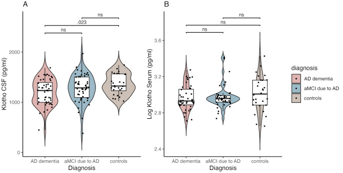
The mean concentration of CSF sαKl was highest in the control group (1359.5 ± 208.1 pg/mL), followed by the aMCI due to AD group (1276.4 ± 227.0 pg/mL), and the AD-dementia group (1196.3 ± 262.4 pg/mL) (Table 2; Fig. 2). ANOVA revealed a significant effect of the study group on CSF sαKl levels (F[2,118] = 3.60, p = .030). Post-hoc test indicated that there was a significant difference between the AD dementia and control group (p = .028) but not between the control and aMCI due to AD group (p = .364) or between the aMCI due to AD and AD dementia group (p = .217). The significant main effect for the study group was explained away when we added age and sex into the analysis (F[2,118] = 1.31, p = .273).
With respect to serum sαKl levels, again, the control group had the highest average sαKl concentration (1129.1 ± 459.0 pg/mL), followed by the aMCI due to AD group (992.3 ± 392.6 pg/mL) and the AD dementia group (962.8 ± 316.9 pg/mL) (Table 2; Fig. 2). The main effect of study group in relation to serum sαKl concentration did not reach statistical significance (F[2, 118] = 1.79, p = .172).

2 ) CSF sαKl concentrations in AD dementia, aMCI due to AD and controls.) Serum sαKl concentrations in AD dementia, aMCI due to AD and controls. Serum sαKl data are presented as log-transformed values. Exact-values are displayed for significant group comparisons; "ns" = non-significant A 2B p
Association between sαKl levels and cognitive performance
In the unadjusted model, a modest positive relationship was observed between CSF sαKl levels and memory (β = 0.19, p = .025) in the entire sample. However, this relationship was attenuated and no longer statistically significant after adjusting the model for age, sex, and years of education (β = 0.08, p = .348). We observed a similar trend for language, which also showed a positive association with CSF sαKl levels (β = 0.24, p = .007), with the association no longer evident after adjusting the model for age, sex, and years of education (β = 0.08, p = .367). We also did not observe an association between CSF sαKl levels and language or memory performance when analyzing the study groups separately or when stratifying the analysis based on APOE ε4 status. Finally, there were no significant associations between CSF sαKl levels with any of the other cognitive domains we investigated after controlling for age, sex, and years of education (all p > .05).
We did not observe any significant associations between serum sαKl levels and memory or any of the other investigated cognitive domains (all p > .05), whether analyzing the entire sample, examining the diagnostic groups separately, or stratifying the analyses by APOE ε4 carrier status. The regression coefficients of each of the linear regression models showing CSF or serum based sαKl in relation to composite cognitive domain z-scores in the entire sample are presented in Supplementary Table 2, Additional File 2. The corresponding stratified models—by diagnostic group and APOE ε4 status—are reported in Supplementary Table 3, Additional File 3.
Discussion
This study aimed to investigate KL-VSHET and sαKl protein levels in relation to diagnostic classification along the AD continuum and cognitive performance. First, we assessed whether KL-VSHET was associated with a reduced likelihood of aMCI or dementia due to AD compared to being cognitively intact and whether this association was different in APOE ε4 carriers compared to non-carriers. Second, we examined KL-VSHET's influence on cognitive performance across the AD continuum, comparing the performance of KL-VSHET carriers and KL-VSNC. Third, we compared levels of serum and CSF sαKl protein in cognitively unimpaired controls, aMCI, and dementia due to AD individuals. Finally, we investigated whether CSF and serum sαKl levels were associated with better cognitive performance across the AD continuum. Through addressing these aims, our study contributes to the growing body of literature by examining domain-specific cognitive performance across biomarker-defined stages of the AD continuum, incorporating both CSF and serum sαKl levels as well as KL-VSHET haplotype. This integrated approach provides a more detailed view of how Klotho may relate to cognitive changes in AD.
We found that KL-VSHET carriers exhibited a 61% lower likelihood of being diagnosed with aMCI due to AD compared to being cognitively unimpaired and that this association was similar in both APOE ε4 carriers and non-carriers. Although the p-value only trended towards significance after controlling for all the covariates, including age, sex, and education (p = .061), the OR was 0.39, which corresponds to Cohen's d of 0.52, a medium effect size. Post-hoc power estimates using G*Power software suggested that we had limited power (power of 0.66) to observe this result as statistically significant, hence predisposing our analyses towards Type II error bias. Further analysis revealed that among individuals with aMCI due to AD, KL-VSHET carriers were 48% less likely to be diagnosed with AD dementia, with a similar trend observed in both APOE ε4 carriers and non-carriers. Although this result did not reach statistical significance (p = .102), the observed effect size (Cohen's d = 0.36) suggests a small-to-moderate association within the aMCI due to AD population. When examining the combined group of individuals with aMCI or dementia due to AD compared to cognitively unimpaired individuals, KL-VSHET carriers showed 41% lower odds of being classified within the cognitively impaired group (p = .205), with a stronger trend within APOE ε4 carriers. The OR of this result was 0.59, corresponding to a Cohen's d of 0.29, a small effect size. Although none of these associations reached statistical significance individually, the consistency of the observed trends suggests that KL-VSHET may be associated with lower odds of aMCI or dementia due to AD, a notion that should be explored further in future research with larger sample sizes and a longitudinal follow-up.
Our results partially align with findings by Belloy and colleagues [11], who also reported a decreased risk of AD in individuals with the KL-VSHET haplotype, but only among APOE ε4 carriers, whereas we observed similar results for APOE ε4 carriers vs. non-carriers. Mechanistically, KL-VSHET might exert its protective effect by reducing the buildup of key AD proteins in the brain, Aβ and phosphorylated tau. Studies have shown lower age- and APOE ε4 -related Aβ and tau burden in cognitively unimpaired KL-VSHET carriers at risk for AD [10, 13, 15, 17, 45]. While some studies found no protective effect of KL-VSHET on Aβ and tau burden in APOE ε4 non-carriers [11, 12, 17], others did observe an effect in this subgroup [14, 15]. Our findings are consistent with the latter, suggesting that KL-VSHET might be associated with lower odds of aMCI and dementia due to AD in both APOE ε4 carriers and non-carriers. This potential spectrum of KL-VSHET influence across APOE ε4 status warrants further exploration with larger samples and more robust study designs to definitively disentangle the nuances of this interaction.
We also found that KL-VSHET carriers in the aMCI due to AD displayed significantly better memory compared to KL-VSNC, even after controlling for age, sex, and education, particularly when we restricted the analyses to APOE ε4 carriers. The same association was not evident in AD dementia individuals. These results may reflect a stage-specific effect of KLOTHO-related mechanisms. Prior studies suggest that KL-VSHET may be most effective during earlier phases of the disease, when amyloid and tau pathology are present but widespread neurodegeneration has not yet occurred [10, 14, 15]. In more advanced stages of AD, neurodegenerative changes may overwhelm any neuroprotective influence conferred by KL-VSHET. Together, these findings suggest that KL-VSHET may (a) help support memory function and (b) serve as a buffer against the deleterious effects of APOE ε4 on memory, specifically in the aMCI stage of AD.
Our results are consistent with those of Neitzel and colleagues, who also found that KL-VSHET was associated with better memory performance. They further noted that the observed association may have been explained by reduced tau burden facilitated by the presence of the effect of KL-VSHET haplotype, especially in APOE ε4 carriers [14].
KL-VSHET did not associate with other cognitive domains, implying its influence might be specific to memory. Previously, some have reported an association between KL-VSHET and better cognitive performance, both in healthy individuals and AD patients [6, 13, 16, 46]. Studies have shown positive associations with specific cognitive domains, such as executive function and memory [13, 46], but also broader global cognition [6, 16]. However, others have found no such association or even results where KL-VSHET reflected poorer cognitive performance [45, 47 –51]. These inconsistencies potentially arise from the moderating influence of age and the complex interplay between KL-VSHET and other biological and epigenetic factors, which demand further investigation. Additionally, it is crucial to consider how KL-VSHET's impact on cognition might differ within the specific context of pathological conditions like AD.
We found no significant differences in CSF or serum sαKl levels between the study groups. In the initial analysis, CSF sαKl levels appeared to differ across the study groups, with controls showing the highest levels, followed by the aMCI due to AD and AD dementia, and a statistically significant difference between controls and AD dementia, resembling the decline observed by Grøndvedt and colleagues [18]. However, while in our analyses, this association was reduced to non-significant after adjusting for age and sex, Grøndvedt et al.'s findings remained significant even after adjustment. Semba and colleagues also found lower CSF sαKl in individuals with AD compared to cognitively unimpaired controls [22], supporting the notion of CSF-specific changes potentially missed by blood measurements. Yet one other study reported higher plasma sαKl l in AD patients, contrary to our serum sαKl findings [25]. Other studies observed no significant changes in plasma sαKl across AD stages [18, 24]. This alignment with our results suggests a potentially limited influence of AD pathology on serum and plasma sαKl levels.
With respect to CSF and serum sαKl in relation to memory and other cognitive domains, we found a tentative association with memory and language overall that was explained again by age, sex, and education. Analyzing individual study groups or stratifying by APOE ε4 status also showed no significant associations between CSF sαKl and memory or other cognitive domains. Similarly, serum sαKl levels lacked significant associations with memory or any other assessed cognitive domains. The weak positive correlation observed between CSF and serum sαKl levels suggests that while there is some degree of systemic-central coupling, sαKl in CSF vs. serum likely reflect distinct biological processes. While serum sαKl is predominantly derived from peripheral sources such as the kidney, CSF sαKl is believed to originate primarily from the choroid plexus, indicating central nervous system-specific production. This divergence may help explain why cognitive associations in our study appeared more pronounced for CSF sαKl compared to serum sαKl, although even the CSF-based findings were modest and did not remain statistically significant after adjustment for demographic variables. Together, these results suggest that while sαKl might be involved in AD pathology, its relationship with memory, at least as measured in this study, appears muted and possibly modulated by demographic factors. These findings contradict previous research where higher levels of sαKl among KL-VSHET carriers were used to explain the results between KL-VSHET and better memory [6]. While animal and human studies show cognitive benefits from elevated sαKl [20, 21, 52 –55], our findings suggest this relationship might be more complex in individuals with AD.
Limitations
This study has several limitations that should be considered. First, the cross-sectional study design does not allow for causal inferences to be drawn. Second, the sample size in this study was relatively small, causing limited statistical power and potentially restricting the generalizability of the findings. Post-hoc power estimates using G*Power software indicated that several comparisons had limited statistical power, including those examining differences in diagnostic classification odds between KL-VSHET and KL-VSNC (power of 0.66 for aMCI, 0.50 for AD dementia), as well as the subsequently stratified analyses by APOE ε4 carrier status (0.11–0.33), indicating a bias towards a Type II error. Therefore, null findings in our study may reflect this bias as much as a true lack of a statistically significant association. In the same context, we did not adjust our results for comparison-wise Type I error, which in our view would exacerbate the existing bias towards Type II error due to lower-than-ideal power. Additionally, KL-VS haplotype and sαKl protein data were not co-collected for all participants, limiting the ability to directly explore genotype-phenotype associations. While we did not assess the relationship between KL-VSHET or sαKl protein and AD biomarkers in this study, these associations were investigated in a prior publication [56]. Finally, although the ELISA kit used in this study demonstrated acceptable intra- and inter-assay variability, the precision of serum sαKl measurements at picogram concentrations may still be limited, given the inherently low abundance of the analyte and potential sensitivity constraints of immunoassay-based detection.
Conclusions
As hypothesized, we observed that carriers of the KL-VSHET haplotype were substantially (although often non-significantly) underrepresented among participants with aMCI due to AD and AD dementia compared to cognitively unimpaired individuals, suggesting a potential association with lower likelihood of cognitive impairment. Contrary to our hypotheses, we found that the association between KL-VSHET and diagnostic group classification was similar for APOE ε4 carriers vs. non-carriers, as opposed to being prominent in APOE ε4 carriers only. In addition, we found that the carriers of KL-VSHET with aMCI due to AD displayed better memory performance compared to those without the haplotype, especially in combination with the risk-increasing APOE ε4, suggesting a buffering effect KL-VSHET against the established negative effect of APOE ε4 on memory. These findings deserve further attention, particularly within longitudinal research and with larger samples.
We did not find consistent evidence for an association between CSF or serum sαKl levels and cognitive status or memory performance in our sample. While previous studies reported associations between sαKl protein and AD pathology, our findings suggest that these relationships might be complex and influenced by demographic factors like age and sex, requiring further research with appropriate control and stratification strategies.
While our findings remain tentative due to limitations, they highlight the potential of KL-VSHET and sαKl protein factor as a target for further investigation and emphasize the need for larger, comprehensive studies. Future research should prioritize longitudinal designs, larger sample sizes, and detailed stratification based on genetic and demographic factors to provide more conclusive evidence.
Supplementary Information
Below is the link to the electronic supplementary material.
Supplementary Material 1
Supplementary Material 2
Supplementary Material 3