
What this is
- () are specialized structures linking the endoplasmic reticulum and mitochondria.
- They play critical roles in lipid synthesis, , and cellular quality control.
- Dysfunction of is implicated in various diseases, including neurodegenerative disorders and metabolic syndromes.
- Emerging therapeutic strategies target MAM pathways to restore cellular function and combat disease.
Essence
- are vital for cellular homeostasis, regulating lipid dynamics and . Their dysfunction contributes to diseases like Alzheimer's and metabolic disorders, highlighting their potential as therapeutic targets.
Key takeaways
- maintain lipid homeostasis and , crucial for mitochondrial function. Disruption of can reduce phosphatidylethanolamine levels by up to 30% and impair ATP production by up to 40%.
- MAM dysfunction is linked to neurodegenerative diseases, with evidence showing that narrow MAM contacts increase amyloid beta production in Alzheimer's disease. This suggests MAM integrity is critical for neuronal health.
- Therapeutic strategies targeting , such as small molecules and gut microbiota-derived metabolites, show promise in restoring function and improving outcomes in metabolic and neurodegenerative diseases.
Caveats
- The review primarily synthesizes existing literature, which may limit the depth of empirical evidence for specific claims. Future studies are needed to validate therapeutic strategies targeting .
- Research on is still evolving, and the complexity of their interactions with various cellular processes presents challenges in fully understanding their role in health and disease.
Definitions
- Mitochondria-associated membranes (MAMs): Specialized contact sites between the endoplasmic reticulum and mitochondria that facilitate lipid exchange and calcium signaling.
- Calcium signaling: The process by which calcium ions regulate various cellular functions, including metabolism and cell survival.
Simplified
Functions and cellular significance
Lipid transfer and membrane homeostasis
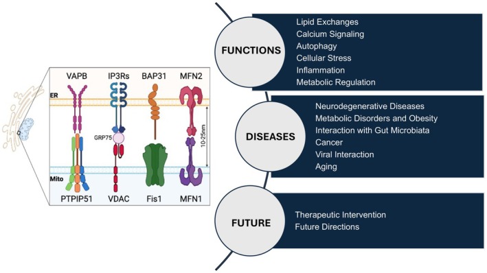
Mitochondria‐associated membranes are specialized contact sites between the ER and mitochondria that support lipid synthesis and interorganelle transfer, crucial for maintaining membrane composition and function [2]. These structures maintain a narrow spacing (10–50 nm) that enables lipid exchange without fusion. Key tethering components form a crucial tripartite complex: the voltage‐dependent anion channel (VDAC1) on mitochondria is physically linked to the ER‐localized inositol 1,4,5‐trisphosphate receptor (IP3R) through the molecular chaperone GRP75, which serves as a bridge between these channels. Additional structural regulators—Mitofusin 2 (MFN2), VAPB, PTPIP51, and PACS2—maintain MAM stability and organization [9, 17].
MAMs are enriched in enzymes that synthesize phosphatidylserine (PS), which is transferred to mitochondria and converted to phosphatidylethanolamine (PE), contributing ~45% of mitochondrial PE. Mitochondria also return newly synthesized PE to the ER, where it serves as a precursor for phosphatidylcholine (PC) synthesis. Cardiolipin, synthesized exclusively in mitochondria, is trafficked to peroxisomes and lysosomes to influence membrane dynamics and function [18]. MAMs also mediate the exchange of PC, cholesterol, and sphingolipids, and regulate cardiolipin synthesis—a mitochondria‐specific phospholipid critical for energy production [9, 19, 20, 21, 22].
These lipid transfers regulate membrane fluidity, protein distribution, and respiratory complex organization. Cardiolipin enrichment, for instance, supports the assembly of respiratory supercomplexes and enhances oxidative phosphorylation [23, 24]. Disruption of MAMs reduces PE levels by up to 30%, mislocalizes cardiolipin, disrupts electron transport, impairs ATP production by up to 40%, and leads to mitochondrial fragmentation [25, 26].
MAMs also coordinate lipid droplet formation by linking ER lipid synthesis to storage pathways. Their disruption reduces lipid droplet formation by ~60%, exacerbating cellular stress and lipid dysregulation [24, 27].
Given their essential roles in lipid balance, mitochondrial function, and organelle communication, MAMs are emerging as therapeutic targets. Pharmacological modulation of MAM integrity may provide novel strategies for metabolic and neurodegenerative diseases. Understanding MAM‐regulated lipid dynamics is critical for leveraging this potential. While lipid transfer establishes MAM structure, calcium signaling across these contact sites is central to metabolic regulation and cell survival.
Calcium signaling and metabolism
MAMs serve as key regulators of calcium signaling, supporting ATP production, apoptosis, and metabolic stability [28]. Calcium transfer from the ER to mitochondria is mediated by the same IP3R–GRP75–VDAC1 complex [29]. This coupling ensures efficient calcium flux while avoiding overload [6, 30].
Within mitochondria, calcium acts as a cofactor for enzymes in the tricarboxylic acid cycle and oxidative phosphorylation, directly modulating ATP output [31]. MAM‐controlled calcium flux allows cells to dynamically adjust their bioenergetics. Excessive calcium transfer can open the mitochondrial permeability transition pore (mPTP), initiating apoptosis under stress [32, 33, 34].
Therapeutically, stabilizing the IP3R–GRP75–VDAC1 complex preserves mitochondrial function and cell viability. For example, intranasal insulin maintains this complex in neurons, offering neuroprotective effects [35, 36]. Similarly, Lipin1 enhances cognitive function in diabetic encephalopathy by restoring calcium homeostasis through MAM integrity [37].
MAM calcium signaling dysregulation is linked to cancer, heart disease, and neurodegeneration [38]. Its dual role in metabolism and stress response presents both a pathological vulnerability and a therapeutic opportunity [39]. In addition to regulating energy metabolism, MAMs also coordinate quality control pathways like autophagy and mitophagy.
Autophagy and mitochondrial quality control
MAMs coordinate autophagy and mitophagy—key quality control pathways that clear damaged organelles and proteins, supporting homeostasis [12].
Autophagy eliminates dysfunctional cellular components. MAMs promote this process under stress (e.g., oxidative stress or starvation) by regulating calcium release via the IP3R–GRP75–VDAC1 complex, which is essential for autophagosome biogenesis and maturation [40, 41].
MAMs also support mitophagy—the selective removal of damaged mitochondria. These sites serve as platforms for mitochondrial tagging and autophagosome formation [39, 42]. Their spatial organization ensures selective degradation of impaired mitochondria, preserving healthy ones and maintaining energy production.
In neurodegeneration, MAM fragmentation impairs autophagy. Kulkarni et al. [43] showed that loss of MAM scaffolding reduces autophagic flux in AD and PD. Pharmacological modulation of MAMs may restore autophagy; for instance, metformin enhances autophagic activity by modulating the IP3R–GRP75–VDAC1 tether, increasing cytosolic calcium in hepatocellular carcinoma [44, 45]. Although this was shown in cancer, the mechanism may apply broadly to autophagy‐impaired diseases.
Through their regulation of autophagy and mitophagy, MAMs help cells adapt to stress by removing dysfunctional components and supporting metabolic integrity. This quality control is vital under pathophysiological conditions [46, 47]. MAMs thus integrate stress signaling and degradation pathways to preserve cellular function. In parallel, MAMs serve as key responders to cellular stress, modulating both the UPR and integrated stress response (ISR) to restore homeostasis.
Cellular stress responses
Mitochondria‐associated membranes are central to cellular stress response mechanisms, contributing to both the UPR and the ISR. These pathways are essential for cellular adaptation and survival under challenging conditions, and MAMs play a key role in coordinating these processes.
Unfolded protein response () UPR
When the ER accumulates misfolded or unfolded proteins, the UPR is activated to restore ER homeostasis [48]. MAMs facilitate this response by serving as hubs for calcium and lipid exchange between the ER and mitochondria, regulated by proteins, such as MFN2 and PACS2 [49]. Calcium transfer at MAMs modulates three ER stress sensors—inositol‐requiring enzyme 1 (IRE1), protein kinase R‐like ER kinase (PERK), and activating transcription factor 6 (ATF6)—which initiate signaling pathways that enhance protein folding and reduce ER load [50]. MAM integrity directly influences both the activation and duration of these pathways, and disrupted contacts can prolong ER stress and trigger apoptosis [51].
Integrated stress response () ISR
The ISR is triggered by diverse stressors including nutrient deprivation, viral infection, and ER dysfunction [52]. MAMs influence this pathway through their regulation of PERK, which also links UPR and ISR signaling. Upon activation, PERK phosphorylates eukaryotic initiation factor 2α (eIF2α), reducing global protein synthesis to conserve resources and promote recovery [53]. Calcium and lipid signaling at MAMs modulate PERK activity and thus impact the broader ISR.
Beyond protein folding and translation control, MAMs influence calcium homeostasis, mitochondrial energy metabolism, and lipid dynamics. Dysregulation of MAMs can compromise these processes and lead to cell death.
In summary, MAMs integrate and regulate UPR and ISR signaling, enabling cells to adapt to stress. Their ability to modulate overlapping yet distinct pathways highlights their role in maintaining cellular resilience.
and inflammation MAMs
MAMs are critical regulators of inflammation, particularly through NLRP3 inflammasome activation and calcium‐dependent immune signaling [54, 55]. The NLRP3 inflammasome localizes to MAMs during activation [8], where these contact sites provide platforms for assembling the inflammasome complex and initiating inflammatory responses. Disrupted calcium handling at MAMs alters cytokine production and immune cell behavior [56].
Immune cell activation andregulation NLRP3
In macrophages, MAM‐localized inflammasome activation requires both mitochondrial reactive oxygen species (ROS) and calcium signaling [57]. The spatial architecture of MAMs integrates these signals, facilitating the assembly of inflammasome components and the production of IL‐1β and IL‐18 [58]. Disruption of this coordination can drive excessive inflammation.
Reactive oxygen species () ROS
MAM dysfunction increases mitochondrial ROS, a potent activator of the NLRP3 inflammasome. ROS also enhance NF‐κB activity and promote inflammasome oligomerization [13, 59], creating a self‐reinforcing inflammatory loop.
stress and inflammation ER
As discussed above, ER stress is modulated at MAMs. Unresolved ER stress can escalate inflammation or lead to cell death, linking MAM dysfunction with broader inflammatory signaling [49].
Mitochondrialrelease DNA
Dysfunctional MAMs may promote mitochondrial DNA (mtDNA) leakage into the cytoplasm, which activates innate immunity via the cGAS–STING pathway and contributes to chronic inflammation [60].
The interplay between MAM integrity and inflammation is increasingly relevant in neuroinflammatory, metabolic, and autoimmune diseases. Clarifying these mechanisms may open therapeutic strategies for controlling maladaptive inflammation.
Metabolic regulation and disease
MAMs are key regulators of lipid metabolism and insulin signaling– both essential to energy homeostasis. They mediate phospholipid transfer between the ER and mitochondria, sustaining membrane composition and mitochondrial integrity [23]. Disruption of these exchanges contributes to ectopic lipid accumulation in the liver and muscle, driving metabolic disorders, such as nonalcoholic fatty liver disease (NAFLD) and insulin resistance [15, 61, 62].
MAMs also influence insulin signaling by hosting interactions between insulin receptor substrates and downstream signaling proteins [63]. They modulate calcium flux and impact the activity of Akt, a central insulin‐signaling kinase [64]. In conditions like chronic overnutrition, MAM expansion alters calcium signaling and impairs insulin action [65]. Loss of MAM integrity has been shown to reduce insulin sensitivity by up to 40% in skeletal muscle cells [66].
By orchestrating lipid dynamics, calcium homeostasis, and insulin responsiveness, MAMs stand at the intersection of cellular metabolism and disease. Restoring MAM function may offer a promising strategy for treating obesity, insulin resistance, and related metabolic disorders.
Role in disease and therapeutic implications
Neurodegenerative and neurocognitive disorders
Mitochondria‐associated ER membranes (MAMs) are increasingly recognized as critical regulators in neurodegenerative diseases, such as AD and PD, as well as neurocognitive and psychiatric disorders. The strategic positioning of MAMs at the interface between the ER and mitochondria makes them critical regulators of multiple cellular processes essential for neuronal survival and function.
Classical neurodegeneration (,) AD PD
Disrupted MAMs impair mitochondrial fission and fusion dynamics, contributing to neuronal degeneration [67, 68]. MAM dysfunction is increasingly recognized as a key factor that disrupts cellular homeostasis and leads to neuronal damage [69].
Recent evidence suggests that MAM integrity is compromised early in disease progression, potentially serving as an initiating event rather than merely a consequence of neurodegeneration [70].
Zellmer et al. [71] showed that narrow MAM contacts increase amyloid beta (Aβ) production and reduce mitochondrial motility in 3D AD neural cultures, while Barbuti et al. [72] found that α‐synuclein localizes to MAMs and disrupts phosphatidylserine metabolism, contributing to regional vulnerability in PD. Additionally, MAM disruption impairs mitophagy, as Luo et al. [73] demonstrated, exacerbating neurodegeneration in AD models.
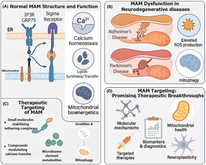
MAMs are central to calcium homeostasis; their dysfunction leads to excessive calcium transfer to mitochondria, resulting in calcium overload, oxidative stress, and neuronal damage [74, 75]. The impact of MAM dysfunction extends beyond mitochondrial dynamics to affect calcium homeostasis, lipid metabolism, and autophagy—all critical processes for maintaining neuronal health. Disruptions in these pathways create a self‐perpetuating cycle of cellular damage that accelerates disease progression [76]. Furthermore, MAM alterations appear to exhibit regional specificity within the brain, potentially explaining the selective vulnerability of certain neuronal populations in different neurodegenerative conditions [77] (Fig. 2).

Mitochondria‐associated membrane (MAM) structure, dysfunction, and therapeutic targeting in neurodegenerative diseases. Comprehensive illustration of MAMs in health, disease, and therapeutic intervention. (A) Normal MAM structure showing key tethering complexes including IP3R–GRP75–VDAC1 and sigma‐1 receptor, which maintain proper calcium homeostasis and lipid transfer between the endoplasmic reticulum and mitochondria. (B) MAM dysfunction in neurodegenerative diseases, depicting disease‐specific alterations including narrowed contacts with increased Aβ production in Alzheimer's disease, α‐synuclein accumulation at MAMs in Parkinson's disease, elevated reactive oxygen species (ROS) production, and impaired mitophagy. (C) Therapeutic targeting of MAMs through multiple approaches including small molecules that stabilize tethering complexes, compounds that modulate calcium transfer, microbiome‐derived metabolites like urolithin A, and interventions enhancing mitophagy and mitochondrial bioenergetics. (D) Emerging therapeutic breakthroughs targeting MAMs, highlighting molecular mechanisms, mitochondrial targets, targeted delivery systems, biomarker development for early diagnosis, and approaches to enhance neuroplasticity through MAM modulation.
Neurocognitive and psychiatric disorders
MAM dysfunction extends beyond classical neurodegenerative diseases to neurocognitive and psychiatric disorders, revealing common pathophysiological mechanisms across neurological conditions. MAMs regulate calcium dynamics in neurons, influencing key processes, such as synaptic plasticity, neurotransmitter release, and neuronal excitability, all of which are essential for learning and memory [77].
Zhang et al. [78] demonstrated in mouse models of depression that chronic stress enhances MAM contact formation and ER–mitochondria calcium transfer in microglia, leading to NLRP3 inflammasome activation and depressive behaviors. This study implicates MAMs in microglial stress responses, further linking MAM dysfunction to neurocognitive deficits.
In schizophrenia, MAM dysfunction contributes to aberrant calcium signaling, mitochondrial impairment, and oxidative stress that underlie the cognitive deficits characteristic of the disorder [79]. Recent investigations have revealed that genetic risk factors for schizophrenia, including DISC1 and NRG1, interact with MAM components, potentially explaining the mitochondrial abnormalities observed in patient‐derived neurons [80].
Alterations in MAM‐mediated mitochondrial function and calcium signaling have been strongly associated with mood disorders, including depression and bipolar disorder [81]. Zhang et al. [78] highlighted that disrupting GRP75, a key protein in MAM formation, reversed stress‐induced MAM changes and depressive behaviors. Yang et al. [82] discussed the potential of natural products targeting MAMs as antidepressant therapies, while studies show that antidepressants can normalize MAM‐associated calcium signaling in preclinical models [83].
Therapeutic approaches and future directions
The recognition of MAM involvement across neurodegenerative and neurocognitive disorders has opened new therapeutic avenues targeting these specialized membrane contacts. Advancements in precision medicine may enable personalized treatments by targeting MAM‐related pathways, improving therapeutic efficacy while minimizing adverse effects. The identification of patient‐specific MAM alterations through advanced imaging and molecular profiling could guide tailored therapeutic strategies.
Research has explored targeting MAM‐associated pathways to restore calcium signaling and mitochondrial function [84]. In ischemic stroke models, downregulation of the VAPB‐PTPIP51 tether reduced MAM integrity, but activation of the PI3K pathway reversed these effects [85]. Etxebeste‐Mitxeltorena et al. [86] identified small molecules that enhance MAM contact, restoring lipid metabolism in ALS patient cells and improving mitochondrial morphology.
Emerging technologies such as CRISPR‐based approaches offer unprecedented opportunities to correct specific genetic defects affecting MAM function [87]. Small molecule modulators of MAM integrity and function are being developed as potential disease‐modifying agents that could be selected based on individual patient profiles. Advanced imaging technologies, including super‐resolution microscopy and cryo‐electron tomography, are deepening our understanding of MAM structure and function with unprecedented detail [88]. Zellmer et al. [89] developed quantitative tools using live‐cell imaging to measure MAM dynamics in 3D neural models, providing methods to assess MAM‐targeting therapeutics.
Biomarker development focused on MAM dysfunction may facilitate early intervention before irreversible neuronal damage occurs. Circulating markers of MAM stress could potentially be detected in blood or cerebrospinal fluid, enabling noninvasive monitoring of disease progression and treatment response [90]. Multi‐omics approaches integrating proteomics, lipidomics, and metabolomics may identify MAM‐associated signatures that predict disease onset and progression.
Metabolic disorders and obesity
MAMs are essential regulators of cellular metabolism, particularly in lipid homeostasis. Their disruption contributes to metabolic syndromes, such as obesity, insulin resistance, and NAFLD, largely through impaired mitophagy, defective mitochondrial quality control, and increased oxidative stress [15, 91].
Therapeutic strategies that target MAMs to enhance calcium signaling and lipid exchange are emerging. Preclinical studies indicate that restoring MAM function can improve metabolic balance and insulin sensitivity in obesity and diabetes models [61, 92]. Drug discovery efforts focused on MAM—associated proteins and lipids are yielding promising candidates for clinical development, including compounds that stabilize MAM contacts, enhance calcium buffering capacity, or improve mitochondrial quality control [93].
Interaction with gut microbiota
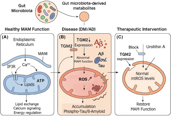
Recent research has uncovered links between gut microbiota and MAM function, providing a new dimension to host metabolic regulation [94]. Microbial metabolites, notably urolithin A produced from ellagitannins in the gut, modulate MAM‐related processes, such as mitochondrial calcium influx and ROS generation. Urolithin A reduces transglutaminase type 2 (TGM2) expression and MAM formation, thereby attenuating mtROS accumulation, suppressing Aβ‐producing enzymes, and improving cognitive outcomes in diabetic models (Fig. 3).
Investigating the interplay between MAMs and the gut microbiota may reveal new insights into disease pathology [94]. The discovery that microbial metabolites can influence MAM function opens exciting possibilities for dietary and probiotic interventions to modulate neurodegeneration and metabolic dysfunction.

Gut microbiota‐derived metabolites modulate mitochondria‐associated membrane (MAM) function in neurodegenerative and metabolic disorders. Schematic illustration of the interplay between gut microbiota, MAMs, and neurodegeneration. (A) In healthy conditions, gut microbiota‐derived metabolites support normal MAM function, facilitating proper calcium signaling, lipid exchange, and energy regulation between the endoplasmic reticulum (ER) and mitochondria via the IP3R–GRP75–VDAC1 complex. (B) In disease states such as diabetes mellitus (DM) and Alzheimer's disease (AD), increased transglutaminase type 2 (TGM2) expression disrupts MAM integrity, leading to elevated mitochondrial reactive oxygen species (ROS) production, abnormal calcium handling, and accumulation of pathological proteins (phospho‐tau and β‐amyloid). (C) Therapeutic intervention with urolithin A, a gut microbiota‐derived metabolite from ellagitannins, blocks TGM2 expression, normalizes mitochondrial ROS levels, and restores MAM function, potentially mitigating neurodegenerative processes. This mechanism represents a novel gut–brain axis pathway with implications for both metabolic and neurodegenerative disorders.
Cancer
MAMs contribute to cancer pathogenesis by facilitating metabolic reprogramming to support uncontrolled cell proliferation [95, 96]. They enhance energy production and biosynthesis, while also modulating calcium signaling and mitochondrial membrane potential to promote apoptosis resistance [97].
Targeting MAM‐associated pathways is emerging as a novel cancer treatment approach. Efforts include disrupting MAM‐mediated metabolic advantages and improving drug sensitivity [98, 99, 100, 101]. Advances in imaging technologies now allow high‐resolution visualization of MAMs, facilitating deeper insight into their roles in tumor biology [102, 103].
Viral interactions and emerging threats
Since 2010, MAMs have been identified as key sites exploited by viruses to facilitate replication and immune evasion. Viruses such as hepatitis C and dengue manipulate MAM‐associated lipid and calcium transfer for replication [104, 105]. HIV‐1 hijacks MAMs at multiple stages of its life cycle, altering calcium and lipid dynamics to enhance viral assembly and budding [16, 106].
The COVID‐19 pandemic and other emerging pathogens like Zika virus have renewed interest in how viruses exploit MAMs [107, 108]. The sigma‐1 receptor (Sig‐1R), a chaperone protein enriched at MAMs, has emerged as a critical factor in viral infections, including SARS‐CoV‐2, making it an attractive target for broad‐spectrum antiviral development.
Aging and senescence
Aging is a major risk factor for neurodegeneration, and age‐related MAM dysfunction contributes to impaired mitochondrial efficiency, calcium dysregulation, and oxidative stress [109, 110, 111, 112]. These deficits promote cellular senescence and inflammation, hallmarks of aging. Wang et al. [113] reported MAM alterations during aging, including disrupted ER–mitochondrial morphology and signaling.
MAMs regulate senescence‐associated mitochondrial function, with proteins like VAPB‐PTPIP51 playing key roles in aging‐linked decline [114, 115]. Targeting MAMs could slow age‐related cellular decline and improve resilience, representing a potential antiaging therapeutic strategy.
Conclusion and future directions
MAMs serve as central orchestrators of cellular homeostasis, linking mitochondrial function, calcium signaling, and lipid metabolism to diverse pathological processes across neurodegenerative, metabolic, infectious, and neoplastic diseases. The emerging understanding of MAM dysfunction reveals common pathophysiological mechanisms that span seemingly distinct conditions, from AD to metabolic disorders to viral infections.
As research continues to elucidate the molecular details of MAM regulation, novel therapeutic strategies targeting MAM integrity and signaling hold promise for addressing complex disease challenges. The convergence of advanced imaging technologies with targeted pharmacological approaches may soon enable precision medicine approaches that restore MAM function in patient‐specific contexts. By targeting the fundamental cellular processes regulated by MAMs, future therapeutic strategies may achieve true disease modification rather than merely symptomatic relief, potentially revolutionizing treatment paradigms across multiple conditions and moving us closer to transformative advances in diagnosing, treating, and ultimately preventing these devastating diseases.
Future research on MAMs will further elucidate their role in cellular communication, disease mechanisms, and therapeutic strategies. Key areas of focus included the following:Molecular mechanisms: Advanced imaging technologies, including super‐resolution microscopy and cryo‐electron tomography, will deepen our understanding of MAM structure and function in health and disease. These approaches will reveal the dynamic nature of MAM contacts and their regulation with unprecedented detail [88].Biomarkers and diagnostics: Continued efforts in biomarker discovery will facilitate early disease detection and treatment monitoring [116]. Multi‐omics approaches integrating proteomics, lipidomics, and metabolomics may identify MAM‐associated signatures that predict disease onset and progression.Targeted therapies: Drug discovery efforts focused on MAM‐associated proteins and lipids are yielding promising candidates for clinical development [93]. These include compounds that stabilize MAM contacts, enhance calcium buffering capacity, or improve mitochondrial quality control.Gut–brain axis: The discovery that microbial metabolites can influence MAM function opens exciting possibilities for dietary and probiotic interventions to modulate neurodegeneration and metabolic dysfunction [94].Neurocognitive function: Understanding how MAMs contribute to synaptic plasticity and neuronal excitability could lead to novel cognitive enhancement strategies and deeper insights into learning and memory mechanisms [79].
As research progresses, MAMs are poised to bridge critical gaps in our understanding, offering new perspectives on disease prevention and treatment. The convergence of advanced technologies will accelerate discoveries in this rapidly evolving field, potentially achieving true disease modification rather than merely symptomatic relief.
Conflict of interest
The authors declare no conflict of interest.
Authors contributions
VB, MS, NS, and BES contributed to manuscript editing. All authors approved the submitted version.
Declaration of generative AI and AI‐assisted technologies
During the preparation of this work, the author(s) used ChatGPT in order to improve clarity, flow, and readability. AI was used exclusively for text editing and refinement, with no involvement in data analysis. After using this tool/service, the author(s) reviewed and edited the content as needed and take(s) full responsibility for the content of the publication.