
What this is
- This research investigates the role of Mtch in during Drosophila intestinal development.
- , the process of clearing damaged mitochondria, is crucial for cellular health.
- The study identifies Mtch as a key regulator that works alongside Vps13D and other proteins like PINK1 and Parkin.
Essence
- Mtch regulates mitochondrial clearance during intestinal development in Drosophila, functioning in a pathway with Vps13D, PINK1, Parkin, and BNIP3.
Key takeaways
- Mtch and Vps13D operate in the same genetic pathway for . Loss of Mtch leads to mitochondrial accumulation, similar to Vps13D mutants.
- defects in Mtch mutants are characterized by elevated levels of autophagy markers like Ref(2)p and Atg8a, indicating impaired mitochondrial clearance.
- Mtch is essential for the proper expression and localization of BNIP3, a receptor, suggesting its role as a mitochondrial protein insertase.
Caveats
- The study primarily uses Drosophila models, which may limit the direct applicability of findings to other organisms, including humans.
- The investigation focuses on developmental stages, which may not reflect processes in adult tissues.
Definitions
- mitophagy: A selective form of autophagy that targets and degrades damaged mitochondria to maintain cellular health.
Simplified
Introduction
Macroautophagy (self-eating, and hereafter autophagy) is an important cytoplasmic recycling process that is critical for cell and organism health [1]. During autophagy, cytoplasmic cargoes are enveloped in an autophagosome and trafficked to lysosomes for degradation [2]. Autophagy can either clear nonspecific cargoes or selectively clear specific cargoes, including the organelles mitochondria, endoplasmic reticulum (ER), and peroxisomes [3]. Alterations in mitochondrial-selective autophagy (mitophagy) have been associated with multiple diseases, including Parkinson's, Alzheimer's, and other diseases [4]. Two of the most frequently mutated genes in familial Parkinson's disease are PINK1 and Parkin, and these two genes are key regulators of mitophagy [5]. Interestingly, PINK1, Parkin, and the mitophagy receptor BNIP3 are each required for the removal of mitochondria in the Drosophila intestine [6,7], but other studies suggest that they function in distinct mitophagy pathways [8]. Thus, it is important to understand the relationship between PINK1, Parkin, and BNIP3 function in mitophagy, and determine how they integrate to clear mitochondria in the physiological setting of the Drosophila intestine during development.
Vps13D, an evolutionarily conserved lipid transfer and ubiquitin binding protein, is required for mitochondrial clearance during Drosophila intestine development and has been implicated in a pediatric movement disorder [9 –11]. Structural studies of Vps13D have revealed that it functions as a lipid conduit between membranes, where a hydrophobic groove acts as a bridge for lipid supply [12]. Vps13D regulates mitochondrial and ER contacts, where it physically interacts with the ER scramblase Vmp1 [13]. Thus, investigating how Vps13D functions at the mitochondrial membrane is essential to understanding its function during mitophagy. Importantly, proteomics analysis revealed that Vps13D physically interacts with multiple outer mitochondrial membrane proteins [14].
MTCH2 is an outer mitochondrial membrane protein with multiple roles in cell health. Originally defined as a regulator of cell death, MTCH2 was characterized to be important in tBID-mediated apoptosis [15]. MTCH2 has also been studied in the context of lipid metabolism, where it is an important regulator of de novo lipogenesis and mitochondrial fusion [16]. Most recently, MTCH2 was described to function in tail-anchored protein insertion into the outer mitochondrial membrane as a mitochondrial protein insertase [17]. Despite extensive investigation, it remains unclear how MTCH2 can play multiple roles in mitochondrial and cell health.
MTCH2 physically interacts with VPS13D in human cells [14]. This prompted our investigation of the relationship between Vps13D and the MTCH2 orthologue Mtch in Drosophila. Here, we identify Mtch as a regulator of mitochondrial clearance during Drosophila intestine development. We show that Mtch and Vps13D function in the same genetic pathway to regulate mitophagy. Importantly, Mtch also functions in a pathway with PINK1, Parkin, and BNIP3. Our data support a model with Mtch functioning as a BNIP3 insertase that is required for mitophagy downstream of PINK1 and Parkin, and this insertase function can potentially explain the diverse cellular functions of MTCH family proteins.
Results
Mtch regulates mitochondrial clearance by autophagy during development
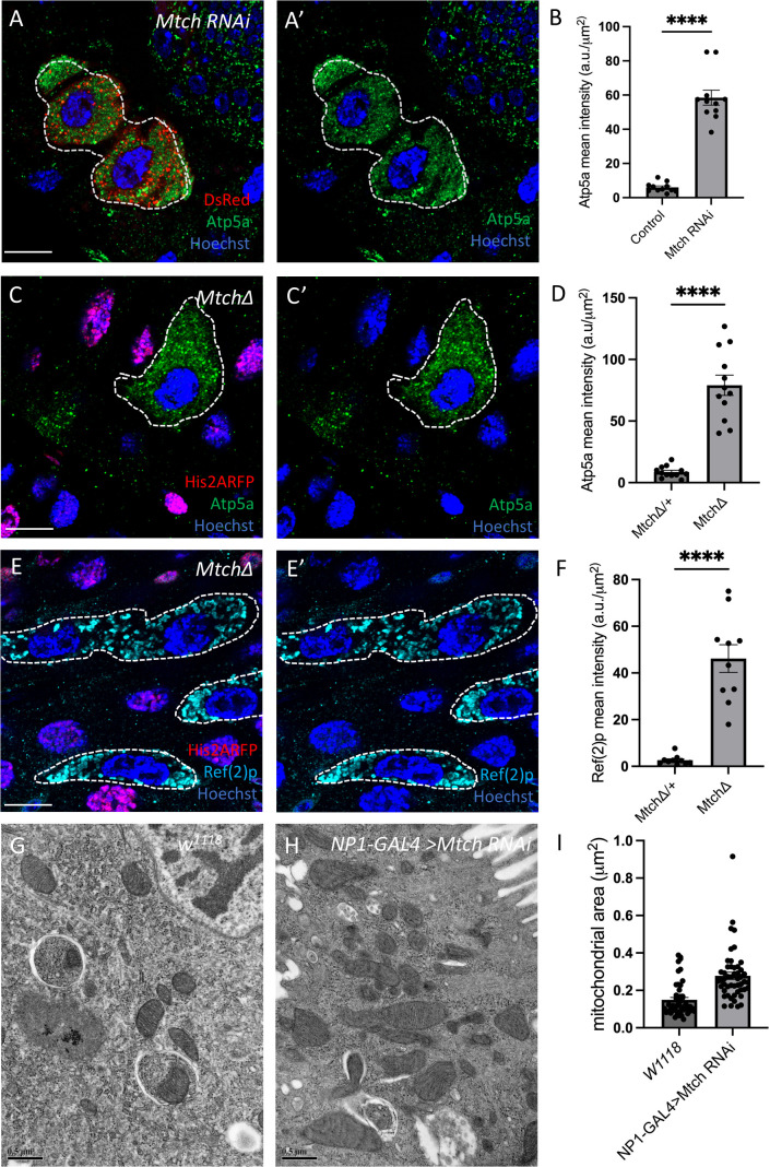
Vps13D is required for the clearance of mitochondria in the developing Drosophila intestine, but the proteins on the mitochondrial surface that interact with and have similar function to Vps13D are poorly understood. Therefore, we screened a group of genes known to encode mitochondrial proteins that physically interact with VPS13D in human cells for phenotypes that are similar to Vps13D in enterocytes of the Drosophila intestine (S1 Table [14]). We examined markers of autophagy and mitochondrial clearance defects, including Ref(2)p, Atg8a, and Atp5a accumulation, in cells expressing RNAi to knockdown gene function 2 h after puparium formation (2 h APF) at a stage soon after the induction of mitophagy. The most robust defects in mitochondrial Atp5a clearance were observed in Mtch RNAi knockdown cells that are marked by the presence of red fluorescent protein (RFP) compared to neighboring control cells (Fig 1A, 1B). In addition, Mtch RNAi knockdown cells also exhibited accumulations of the autophagy adaptor protein Ref(2)p and core autophagy protein Atg8a compared to control neighboring cells (S1A–S1D Fig). Importantly, a second independent RNAi strain that targets Mtch resulted in the same accumulation of Atp5a and Ref(2)p in intestine enterocytes (S1E–S1F' Fig). Thus, knockdown of Mtch phenocopies Vps13D mutant defects in enterocytes of the developing midgut. To confirm the Mtch RNAi phenotypes, we used CRISPR-Cas9 gene editing to delete the Mtch open reading frame and generate a null mutant allele. Similar to RNAi knockdown, Mtch null mutant enterocyte cells accumulate Atp5a compared to neighboring control cells 2 h APF (Fig 1C and 1D). Mtch mutant intestine cells also possess elevated cytoplasmic levels of the autophagy substrate Ref(2)p and the core autophagy protein Atg8a (Figs 1E–1H and S1G–S1H). Combined, these data indicate that Mtch mutant cells exhibit similar phenotypes to intestine enterocyte cells lacking Vps13D function.
We next investigated the function of Mtch in mitophagy by expressing the tandem GFP-RFP Mito-QC transgene in intestine enterocytes. The Mito-QC construct contains a portion of the mitochondrial tail-anchored protein Fis1 that enables association with mitochondria so that mitochondrial clearance can be followed during mitophagy. When autophagosomes fuse with acidic lysosomes pH-sensitive GFP signal is lost, and when mitophagy is dysfunctional GFP persists. Importantly, RFP signal is present in both autophagosomes and acidic autolysosomes. We tested Mito-QC in Mtch mutant cells and found that both GFP and RFP signal were decreased compared to control neighboring cells even when the signal in control cells was saturated (S2A–S2A" Fig). We also used a Mito-GFP construct that is associated with mitochondria because of mitochondrial COX8 sequence, and observed a loss of GFP in Mtch mutant cells compared to control neighboring cells (S2B–S2B' Fig). These results prompted analysis of the influence of Mtch on mitochondria using Transmission Electron Microscopy (TEM) in intestine enterocytes 2 h APF. Homozygous mutant Mtch animals are lethal during development. Thus, we expressed Mtch RNAi in the enterocytes of the intestine. Mtch knockdown intestine cells exhibited increased numbers of mitochondria compared to control w1118 cells (Fig 1G–1I). Consistent with both TEM and Atp5a immuno-staining data, intestine enterocytes with reduced Mtch function possess both elevated COX4 antigen and more mitochondria that possess membrane potential based on TMRE staining (S2C–S2F Fig). In addition, Mtch mutant intestine cells also exhibited equivalent amounts of cleaved caspase-3 staining as neighboring control cells (S2G–S2G' Fig), suggesting that caspases are not causing mitochondrial content differences. These data reveal that widely used mitophagy sensors may not always provide an accurate reflection of mitochondrial clearance, as Atp5a, COX4, TEM, and TMRE analyses revealed the presence of mitochondria in Mtch mutant cells. In addition, these data reveal that Mtch has a previously unknown role in mitochondrial clearance, and that Mtch loss-of-function phenotypes in the intestine enterocytes are similar to Vps13D mutant cells.

is required for mitochondrial clearance. Mtch (A and A')RNAi (v106996) knockdown cells (DsRed labeled, white dotted outline) accumulate the mitochondrial protein Atp5a (green). Scale bar is 20 μm.Quantification of Atp5a fluorescence intensity inknockdown and control cells ( = 11and = 11 control cells across 3 independent animals were measured). Data are presented as mean ± SEM. **** < 0.0001. (C and C')mutant cells (absence of nuclear RFP, white dotted outline) accumulate the mitochondrial protein Atp5a (green). Scale bar is 20 μm.Quantification of Atp5a fluorescence intensity in mutant and control cells ( = 12and = 12 control cells across 3 independent animals were measured). Data are presented as mean ± SEM. **** < 0.0001. (E and E')mutant cells (absence of nuclear RFP, white dotted outline) accumulate autophagy adaptor Ref(2)p (cyan). Scale bar is 20 μm.Quantification of Ref(2)p fluorescence intensity inmutant and control cells ( = 10 knockout and = 10 control cells across 3 independent animals were measured). Data are presented as mean ± SEM. *** < 0.001.Electron micrograph ofcontrol enterocyte in theintestine 2 h after puparium formation. Scale bar is 0.5 μm.Electron micrograph of enterocyte cell-specificRNAi in theintestine at 2 h after puparium formation. Scale bar is 0.5 μm.Quantification of mitochondrial area ofknockdown intestine cells compared to control. ( = 50 mitochondria across 3 separate experiments). Data are presented as mean ± SEM. **** < 0.0001. The underlying data can be found in. Mtch Mtch n Mtch n p Mtch n Mtch n p Mtch Mtch n n p w Drosophila Mtch Drosophila Mtch n p (B) (D) (F) (G) (H) (I) 1118 S1 Data
Mtch regulates Vps13D-dependent mitophagy
We next investigated the relationship between Mtch and Vps13D during developmentally programmed mitophagy in Drosophila intestine enterocytes. Antibodies do not exist to detect Mtch in Drosophila. Therefore, CRISPR-Cas9 gene editing was used to make an in-frame insertion of a 2x-HA epitope with the Mtch open reading frame on the C-terminus of the protein (S3A Fig). Mtch-2xHA is expressed in enterocytes of the intestine, and co-localizes with mitochondrial Atp5a (S3B–S3B" Fig). We confirmed that knockdown of Mtch by expression of RNAi in DsRed-expressing enterocytes significantly reduced Mtch-2xHA levels compared to neighboring control cells (S3C, S3D Fig). Interestingly, high resolution analysis of Mtch and a previously characterized endogenously tagged Vps13D-3xFLAG [13] revealed limited co-localization of these proteins (S3E Fig). Importantly, we observed that Mtch localizes in rings that are likely the outer mitochondrial membranes, and that multiple points of contact exist with Vps13D puncta with substantial lack of co-localization (S3F and S3G Fig). These results suggest that the relationship between Mtch and Vps13D may be dynamic, and raise questions about their direct relationship in a mitophagy pathway.
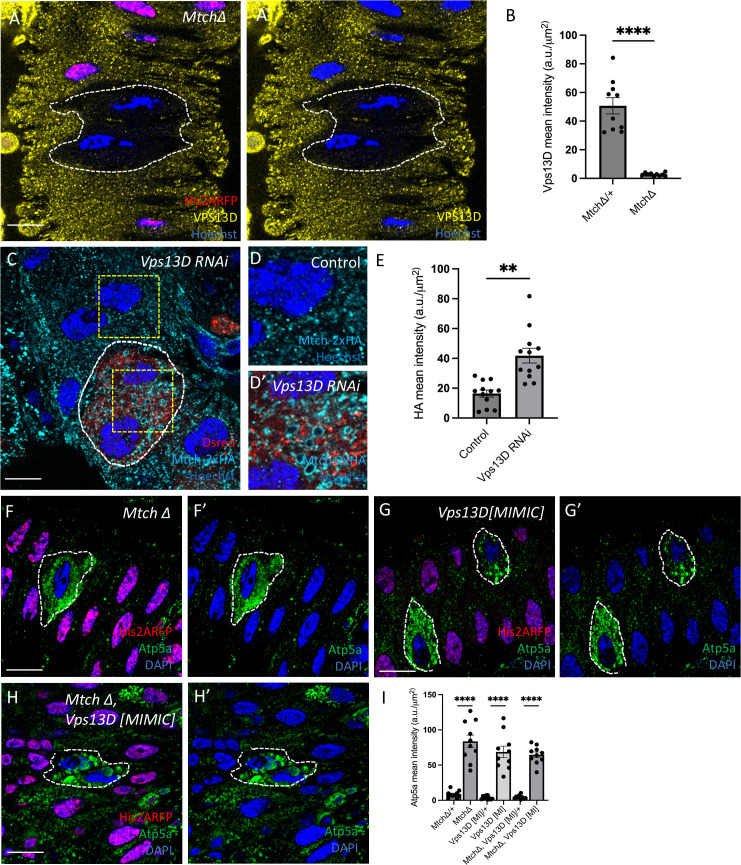
The similarity of Mtch and Vps13D phenotypes, despite limited co-localization (S3F and S3G Fig), prompted us to investigate if their loss influences the levels and localization of one another. We examined the influence of Mtch loss-of-function mutant cells that were stained with an antibody against Vps13D [9]. Vps13D protein puncta levels decreased dramatically in Mtch mutant cells compared to control neighboring intestine cells 2 h APF (Fig 2A, 2B). Importantly, Vps13D RNA levels are similar in intestine enterocytes of control and Mtch knockdown (S3H Fig), indicating that the change in Vps13D is at the level of the protein. Reciprocally, Vps13D function was reduced by expression of RNAi in cells that were marked with DsRed and stained for Mtch-2xHA. Mtch levels were slightly increased in Vps13D knockdown compared to neighboring control intestine cells (Fig 2C–2E). In addition, Vps13D knockdown cells exhibit large rings of Mtch compared to control neighboring cells (Fig 2D and 2D'). These data indicate that Mtch is required for proper Vps13D protein levels, and that Vps13D also influences Mtch abundance and distribution in enterocytes.
To further explore the genetic relationship between Mtch and Vps13D, we performed single and double mutant analysis to compare the levels of mitochondrial Atp5a and the autophagy protein Atg8a in enterocytes. Homozygous Mtch and Vps13D single mutant cells each exhibited elevated levels of Atp5a compared to neighboring control enterocytes 2 h APF (Fig 2F, 2G, and 2I). In addition, Mtch Vps13D double mutant cells did not exhibit an additive change in Atp5a levels compared to single mutants (Fig 2F–2I), indicating that Mtch and Vps13D function in the same mitochondrial clearance pathway. Analysis of Atg8a in Mtch and Vps13D single and double mutants did not reveal a significant difference in Atg8a levels (S4A–S4D Fig). Combined, these data indicate that Vps13D and Mtch function in the same mitophagy pathway.

Relationship between Mtch and Vps13D during mitophagy. (A and A')mutant cells (absence of nuclear RFP, white dotted outline) have decreased levels of Vps13D protein (yellow) in the developingintestine. Scale bar is 20 μm.Quantification of Vps13D fluorescence intensity in mutant and control cells ( = 10mutant and = 10 control cells across 3 independent animals were measured). Data are presented as mean ± SEM. **** < 0.0001.RNAi knockdown cells (Dsred labeled, dotted line) have increased Mtch-2xHA levels (cyan). Scale bar is 20 μm.Enlargement of Mtch-2xHA in control cell. Scale bar is 5 μm. (D') Enlargement of Mtch-2xHA levels inknockdown cell showing large rings of Mtch compared to control (D).Quantification of Mtch-2xHA fluorescence intensity inknockdown and control cells ( = 12knockdown and = 12 control cells across 3 independent animals were measured). Data are presented as mean ± SEM. ** < 0.01. (F and F')mutant cells (absence of nuclear RFP labeled, white dotted outline) inhibit clearance of mitochondrial protein Atp5a (green) compared to neighboring controls (nuclear RFP labeled). Scale bar is 20 μm. (G and G')mutant cells (absence of nuclear RFP labeled, dotted line) inhibit clearance of mitochondrial protein Atp5a (green) compared to neighboring controls (nuclear RFP labeled). Scale bar is 20 μm. (H and H')double mutant cells (absence of nuclear RFP labeled, dotted line) inhibit clearance of mitochondrial protein Atp5a (green) compared to neighboring control cells (nuclear RFP labeled). Scale bar is 20 μm.Quantification of Atp5a mean fluorescence intensity in, andknockout cells ( = 10 for each single and double mutant, and = 10 control cells across 3 independent animals were measured). Data are presented as mean ± SEM. **** < 0.0001. The underlying data can be found in. Mtch Drosophila n Mtch n p Vps13D Vps13D Vps13D n Vps13D n p Mtch Vps13D Mtch Vps13D Mtch, Vps13D Mtch Vps13D n n p (B) (C) (D) (E) (I) S1 Data
Mtch functions in a pathway with PINK1, Parkin, and BNIP3
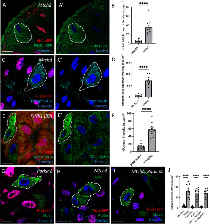
Our data indicates that Mtch and Vps13D function in the same mitophagy pathway. Thus, we next investigated the relationship between Mtch and previously known regulators of mitophagy in the Drosophila intestine during development, including PINK1, Parkin, and the mitophagy receptor BNIP3. Interestingly, PINK1 levels were significantly increased in Mtch mutant enterocytes compared to neighboring control cells 2 h APF (Fig 3A, 3B). Pink1 is required for phosphorylation of ubiquitin and clearance of mitochondria in enterocytes [7]. Consistent with elevated PINK1 levels, we detected significantly increased levels of phosphorylated ubiquitin in Mtch mutant intestine cells compared to control enterocytes (Fig 3C, 3D). We also observed increased Mtch protein levels in PINK1 mutant compared to control intestine cells 2 h APF (Fig 3E, 3F), likely because of the failure to clear mitochondria in Pink1 mutant cells. These data suggest Mtch and Pink1 function in the same mitophagy pathway.
Pink1 phosphorylates both ubiquitin and Parkin to regulate mitophagy [5], and Parkin is required for proper mitophagy in Drosophila intestine enterocytes [7]. Therefore, we investigated the relationship between Mtch and Parkin during mitophagy in the developing intestine. Importantly, single and double mutant analysis revealed similar elevated levels of mitochondrial Atp5a in Mtch and Parkin single mutants, as well as in Mtch Parkin double mutants, compared to neighboring control enterocytes at 2 h APF (Fig 3G–3J). In addition, Mtch levels were elevated in Parkin RNAi knockdown cells compared to neighboring control enterocytes at 2 h APF (S5A, S5B Fig), and this is likely because mitochondrial clearance is inhibited in cells with reduced Parkin function. These data support a model with Mtch and Parkin functioning in the same genetic pathway to regulate mitophagy in developing enterocytes.
BNIP3 encodes a mitophagy receptor that has been reported to function in a PINK1- and Parkin-dependent mitophagy pathway [18,19], and BNIP3 is required for mitophagy in Drosophila enterocytes where PINK1 and Parkin regulate mitochondrial clearance [7]. This prompted us to investigate the relationship between Mtch and BNIP3 during intestine mitophagy. We compared single and double mutants to determine if Mtch and BNIP3 function in the same mitophagy pathway. These experiments revealed similar elevated levels of mitochondrial Atp5a in Mtch and BNIP3 single and in Mtch BNIP3 double mutants compared to neighboring control enterocytes at 2 h APF (Fig 4A–4D). These data indicate that Mtch and BNIP3 function in the same genetic pathway to control enterocyte mitophagy during intestine development.
The Mtch orthologue MTCH2 has diverse functions in apoptosis, lipid metabolism and other processes [15,16,20]. MTCH2 has been reported to function as an insertase that is required for insertion of tail-anchored proteins into the mitochondrial outer membrane [17]. Since BNIP3 encodes a tail-anchored mitophagy receptor, we hypothesized that the function of Mtch in Vps13D-, PINK1-, and Parkin-dependent mitophagy may be through the proper regulation and localization of BNIP3 in the mitochondrial membrane. We utilized a strain of Drosophila that contains a stable human BNIP3 (hBNIP3) with an HA tagged transgene to test this hypothesis. We first expressed hBNIP3 in Drosophila intestine enterocytes, and observed similar low levels of Atp5a in BNIP3 mutant and neighboring control cells (S6A, S6B Fig), indicating that hBNIP3 rescues mitophagy deficiency in BNIP3 mutant enterocytes. By contrast, expression of hBNIP3 in Mtch mutant intestine enterocytes that lack green fluorescent protein (GFP) failed to rescue clearance of mitochondria based on elevated levels of mitochondrial Atp5a in Mtch mutant cells compared to lower levels of Atp5a in neighboring control cells (Fig 4E, 4F). In addition, we used immuno-staining of intestines with Mtch, Vps13D, and Parkin mutant cells that lack GFP at 2 h APF to interrogate expression and localization of hBNIP3. Although hBNIP3 exhibited rings of localization with mitochondria in Vps13D mutant cells (Fig 4G–4I), Mtch mutant enterocytes exhibited lower levels of diffuse hBNIP3 localization that was similar to neighboring control cells (Fig 4J, 4K). Similar to Vps13D mutant cells, Parkin mutant cells accumulated hBNIP3 and had elevated Atp5a (S6C, S6D Fig). Furthermore, when we expressed hBNIP3 in the intestines of Mtch and Vps13D double mutants, we saw decreased levels of hBNIP3, suggesting that Mtch is necessary for BNIP3 localization (Fig 4L, 4M). e additionally tested how hBNIP3 levels change in Mtch and Parkin double mutants and observed decreased levels of hBNIP3 (S6F, S6G Fig). These results indicate that Mtch is required for hBNIP3 localization to mitochondria and mitophagy, and suggest that the influence of Mtch on mitochondrial clearance is because of impaired tail-anchored protein insertion in mitochondria. We tested this possibility by investigating if Mtch loss influences the levels of the Hid and Zucchini (Zuc) mitochondrial tail-anchored proteins. Consistent with our hypothesis, Mtch mutant cells have reduced Hid and Zucchini levels compared to control neighboring cells (S6H–S6J Fig). Combined, these data indicate that Mtch functions in Vps13D-, PINK1-, and Parkin-dependent mitophagy, and that this is likely through the regulation of insertion of the tail-anchored protein and mitophagy receptor BNIP3.

Mtch, PINK1, and Parkin function in a mitophagy pathway. (A and A')mutant cells (absence of nuclear RFP labeled, dotted line) exhibit increased levels of PINK1-GFP (green) compared to control cells (nuclear RFP labeled). Scale bar is 20 μm.Quantification of PINK-GFP mean fluorescence intensity inmutant vs. control cells ( = 10mutant and = 10 control cells across 3 independent animals were measured). Data are presented as mean ± SEM. **** < 0.0001. (C and C')mutant cells (absence of nuclear RFP labeled, dotted line) exhibit increased levels of phosphorylated ubiquitin (cyan) compared to control cells (nuclear RFP labeled). Scale bar is 20 μm.Quantification of phosphorylated ubiquitin mean fluorescence intensity in mutant vs. control cells ( = 10mutant and = 10 control cells across 3 independent animals were measured). Data are presented as mean ± SEM. **** < 0.0001. (E and E')mutant cells (absence of RFP labeled, dotted line) exhibit increased levels of Mtch-2xHA (green) compared to control cells (RFP labeled). Scale bar is 20 μm.Quantification of Mtch-2xHA mean fluorescence intensity in PINK1 in mutant vs. control cells ( = 10 mutant and = 10 control cells across 3 independent animals were measured). Data are presented as mean ± SEM. **** < 0.0001.mutant cells (absence of nuclear RFP labeled, dotted line) inhibit clearance of mitochondrial protein Atp5a (green). Scale bar is 20 μm.mutant cells (absence of nuclear RFP labeled, dotted line) inhibit clearance of mitochondrial protein Atp5a (green). Scale bar is 20 μm.anddouble mutant cells (absence of nuclear RFP labeled, dotted line) inhibit clearance of mitochondrial protein Atp5a (green). Scale bar is 20 μm.Quantification of Atp5a mean fluorescence intensity in mutant vs. control cells ( = 10–12 mutant and = 10–12 control cells across 3 independent animals were measured). Data are presented as mean ± SEM. **** < 0.0001. The underlying data can be found in. Mtch Mtch n Mtch n p Mtch n Mtch n p PINK1 n n p Parkin Mtch Mtch Parkin n n p (B) (D) (F) (G) (H) (I) (J) S1 Data

Mtch and BNIP3 function in a mitophagy pathway. mutant cells (absence of nuclear RFP labeled, dotted line) inhibit clearance of the mitochondrial Atp5a protein (green). Scale bar is 20 μm.mutant cells (absence of nuclear RFP labeled, dotted line) inhibit clearance of the mitochondrial Atp5a protein (green). Scale bar is 20 μm.anddouble mutant cells (absence of nuclear RFP labeled, dotted line) inhibit clearance of the mitochondrial Atp5a protein (green). Scale bar is 20 μm.Quantification of Atp5a mean fluorescence intensity in mutant vs. control cells ( = 10–12 mutant and = 10–12 control cells across 3 independent animals were measured). Data are presented as mean ± SEM. **** < 0.0001. (E and E') Intestines expressing human BNIP3 (hBNIP3) in all enterocytes exhibit elevated levels of Atp5a (purple) inmutant cells (non-GFP labeled, dotted line) compared to neighboring control cells. Scale bar is 20 μm.Quantification of Atp5a mean fluorescence intensity inmutant vs. control cells ( = 11 mutant and = 11 control cells across 3 independent animals were measured). Data are presented as mean ± SEM. **** < 0.0001. (G and G') Intestine cells expressing human BNIP3 (hBNIP3, cyan) in all enterocytes exhibit elevated levels of Atp5a (purple) and rings of hBNIP3 inmutant cells (non-GFP labeled, dotted line) compared to neighboring control cells indicating that hBNIP3 expression fails to rescuemutant mitochondrial clearance. Scale bar is 20 μm.Quantification of BNIP3-HA mean fluorescence intensity inmutant vs. control cells ( = 10 mutant and = 10 control cells across 3 independent animals were measured). Data are presented as mean ± SEM. n.s. = not significant.Quantification of Atp5a mean fluorescence intensity inmutant vs. control cells ( = 10 mutant and = 10 control cells across 3 independent animals were measured). Data are presented as mean ± SEM. n.s. = not significant. (J and J') Intestine cells expressing human BNIP3 (hBNIP3, cyan) in all enterocytes exhibit decreased levels of BNIP3-HA inmutant cells (non-GFP labeled, dotted line) compared to neighboring control cells. Scale bar is 20 μm.Quantification of BNIP3-HA mean fluorescence intensity inmutant vs. control cells ( = 11 mutant and = 11 control cells across 3 independent animals were measured). Data are presented as mean ± SEM. n.s. = not significant. (L and L') Intestine cells expressing human BNIP3 (hBNIP3, cyan) in all enterocytes exhibit decreased levels of BNIP3-HA indouble mutant cells (non-GFP labeled, dotted line) compared to neighboring control cells. Scale bar is 20 μm.Quantification of BNIP3-HA mean fluorescence intensity in, Vps13D double mutants vs. control cells ( = 10 mutant and = 10 control cells across 3 independent animals were measured). Data are presented as mean ± SEM. n.s. = not significant. The underlying data can be found in. (A) (B) (C) (D) (F) (H) (I) (K) (M) BNIP3 Mtch Mtch BNIP3 n n p Mtch Mtch n n p Vps13D Vps13D Vps13D n n Vps13D n n Mtch Mtch n n Mtch, Vps13D Mtch n n S1 Data
Discussion
Mitochondria are key organelles in cellular health that produce ATP as well as having other important functions, such as being signaling scaffolds and key sources of regulators of cell death [21]. Mitochondria are cleared by mitophagy to limit the release of inflammatory factors, including mitochondrial DNA and reactive oxygen species (ROS) [22]. Mitophagy selectively targets and eliminates damaged mitochondria before they can rupture and activate intrinsic apoptosis [23]. Pink1 and Parkin are two of the most studied regulators of mitophagy [24], but alternative Pink1- and Parkin-independent mitophagy pathways exist [6,25]. Thus, it is important to identify additional factors that function in both Pink1/Parkin-dependent and -independent mitophagy pathways.
Using a developmental model to study mitophagy in a physiological setting [26], we discovered that Mtch is required for mitochondrial clearance in the Drosophila intestine. We screened for genes encoding proteins known to interact with Vps13D, a regulator of mitochondrial clearance in the Drosophila intestine, to gain insight into the function of Vps13D during mitophagy. CRISPR-targeted deletion of Mtch revealed strong phenotypes that are similar to Vps13D mutant cells. In addition, Vps13D protein levels are reduced in Mtch mutant cells. Single and double mutant analyses indicate that Mtch and Vps13D function in the same mitochondrial clearance pathway.
PINK1 and Parkin are widely studied because of their key roles in mitochondrial quality control and clearance [5]. We analyzed how Mtch and these two key mitophagy genes influence one another. Interestingly, PINK1 levels were significantly increased in Mtch mutants compared to control cells. PINK1 functions as a kinase to phosphorylate ubiquitin [5], and we observed significant increases in phosphorylated ubiquitin in Mtch cells. These data suggest that PINK1 function is activated in Mtch mutant cells despite a block in mitochondrial clearance by mitophagy. We also observed a dramatic increase in Mtch levels in Pink1 mutants, but this is likely because of the failure of Pink1 mutants to clear mitochondria. Our data also indicate that Mtch and Parkin function in the same mitophagy pathway, as double mutants exhibited similar increases in mitochondria compared to either Parkin or Mtch single mutant intestine cells. Thus, these studies indicate that Mtch functions downstream of Pink1 and Parkin in the regulation of mitophagy in intestine cells.
Mtch/MTCH2 has been reported to possess a variety of functions in diverse taxa, including lipid homeostasis [27], sperm development [28], stem cell maintenance [29,30], mitochondrial fusion [31], apoptosis [15], and most recently as a mitochondrial protein insertase [17]. BNIP3 is required for mitophagy in the Drosophila intestine, and expression of hBNIP3 rescued the BNIP3 mutant mitochondrial clearance defect in intestine enterocytes. In addition, single and double mutant analyses of Mtch and Bnip3 mutants showed similar levels of elevated mitochondrial Atp5a, further indicating that these genes function in a common mitochondrial clearance genetic pathway. Since BNIP3 encodes a mitochondrial tail-anchored protein that acts as a mitophagy receptor [32], we tested the hypothesis that Mtch is required for proper BNIP3 expression and localization with mitochondria. Significantly, hBNIP3 could not rescue clearance of mitochondria in Mtch mutant intestine cells, and hBNIP3 levels were reduced in Mtch mutant cells compared to either Vps13D or Parkin mutant cells. Double mutants of both Mtch and Vps13D as well as Mtch and Parkin both showed reduced levels, which suggests that Mtch is essential for mitophagy despite BNIP3 levels being increased in single mutants of Vps13D and Parkin. In addition, the tail-anchored Mito-QC mitophagy sensor, as well as the mitochondrial tail-anchored proteins Hid and Zucchini, levels were also significantly decreased in Mtch mutant cells. By contrast and similar to Atp5a and Pink1, mitochondrial COX4 protein is increased in Mtch mutant cells, as are levels of TMRE, a reporter of mitochondrial function. Although we have not tested if mitochondrial clearance can be rescued by artificially tethering BNIP3 to mitochondria in Mtch mutant cells, our combined data suggest that BNIP3 mitochondrial localization is necessary for Mtch-mediated mitophagy. Furthermore, our data suggest that the mitochondrial insertase function of Mtch/MTCH2 may explain the broad range of cellular functions attributed to this important mitochondrial protein.
Materials and methods
Fly stocks
Drosophila strains used in this study are listed in the S2 Table with complete genotypes for each experiment in the S3 Table. All crosses were raised at 25 °C. The w1118 wild-type strain was used as control for TEM experiments. Flies were raised on standard cornmeal/molasses/agar media.
Gene editing for deletion of Mtch and tagging of Mtch
The MtchΔ and Mtch-2xHA strains were edited using CRISPR/Cas9 technology [33]. The gRNAs for each strain were cloned into Drosophila-optimized U6b plasmids. For the MtchΔ strain, 0.8kb homology arms were synthesized by IDT and cloned into a TOPO TA vector. For the Mtch-2xHA strain, 1 kb homology arms were synthesized and cloned into a TOPO TA vector. A linker peptide was used between Mtch and the 2xHA peptide, and was designed to be on the C-terminus of Mtch. Plasmid DNA for gRNAs and template were injected into a vasa-cas9 Drosophila germline by Rainbow Genetics. Genotyping was done using primers outside of gRNAs for the MtchΔ strain, while genotyping was done for Mtch-2xHA using primers that included part of the HA sequence and Mtch sequence. Results were then sequenced (Azenta) to confirm the appropriate MtchΔ and Mtch-2xHA sequences. Sequences and gene blocks used are listed in S4 Table.
Induction of mutant and RNAi-directed mosaic cell clones and whole intestine RNAi expression
Mosaic mutant cells and RNAi expression clones were induced as previously described [9]. For RNAi experiments, the RNAi of the gene of interest was crossed to hs-flp;; ACT > CD2 > GAL4, UAS-DsRed line to induce gene knockdown cells. Mutant cell clones (Mtch, Vps13D, Parkin, BNIP3, and respective double mutant combinations) were induced when crossed to a hs-flp;; His2a-RFP, FRT2A strain so that mutant cells were marked by the lack of RFP in the nucleus. For hBNIP3-HA experiments, a hs-flp; NP1-GAL4; Ub-GFP, FRT2A strain was used to induce mutant cell clones. For PINK1[B9] analysis, a hs-flp, FRT19A;; Ub-RFP strain was used to mark mutant cells by the lack of red fluorescence. NP1-GAL4 is an enterocyte-specific GAL4 driver that was used for TEM experiments and to express hBNIP3 and PINK1 RNAi in enterocytes of the entire intestine.
Immunolabeling and microscopy
Crosses were kept in vials with media to mate for 5 h before being subjected to a 1-h heat shock at 37 °C to induce FLP/FRT recombination early in development. Animals were then collected as white prepupae, aged for 2 h at 25 °C on a PBS-soaked Kimwipe, and then promptly dissected in PBS to isolate the intestine. The intestines were then fixed in 4% PFA for 30 min and then incubated in the primary antibodies Ref(2)p (1:200), Atp5a (1:1000), phospho-ubiquitin (1:500), HA (1:500), FLAG (1:500), Atg8a (1:100), Vps13D (1:50), GFP (1:5000), and Hid (1:500). The next day the intestines were washed in 1x PBS 3 times for 15 min before being incubated in secondary antibodies (1:500 for all) for 2 h and Hoechst stained (1:200) for 30 min. Stained intestines were then transferred to microscope slides with Vectashield on them and mounted with a cover slip to be imaged within the next day. Images were taken on a Zeiss LSM700 and LSM900 using a 63x oil objective. At least three animals were imaged for each genotype, with a minimum of 10 cells analyzed per genotype per antibody stain.
For TMRE labeling and imaging, Mtch RNAi was crossed to hsflp;P{y[+t7.7] w[+mC]=CoinFLP-GAL4}attP40 P{w[+mC]=UAS-2xEGFP}AH2 to induce Mtch knockdown cells marked by GFP. Animals were collected as white prepupae, aged for 2 h on a PBS-soaked Kimwipe, and promptly dissected in Schneider's Drosophila Medium (Gibco, 21720024). The intestines were then incubated in the dark with 500 nM TMRE (Invitrogen, T669) diluted in Schneider's medium at room temperature for 20 min. Intestines were immediately mounted in Schneider's medium and imaged on a Zeiss LSM900 using a 63x oil objective. Each intestine was mounted separately, and one image was taken per intestine to reduce variability due to mechanical pressure on the intestine from mounting.
RNA isolation and RT-qPCR analysis
Total RNA was extracted from 12 to 17 pooled intestines per replicate of staged white prepupae using RNeasy Plus Mini Kit (QIAGEN, 74134) and cDNA was synthesized by Super Script III First-Strand Kit (Invitrogen, 18080051) according to the manufacturer's guidelines. Quantitative PCR (qPCR) was performed in triplicate with SYBR Green PCR Master Mix (Applied Biosystems, 4367659) using CFX96 Touch Real-Time PCR Detection System (Bio-Rad, CA, USA). The RNA levels of Vps13D were normalized to RPL32 and are presented as means ± SEM. Primers sequences are listed in S4 Table.
Transmission electron microscopy
Intestines were dissected from animals in PBS 2 h after pupariation. Intestines were then fixed overnight at 4 °C in 2.5% glutaraldehyde and 2% paraformaldehyde in 0.1 M sodium cacodylate buffer, pH 7.4. Tissue was then osmicated and washed in distilled water. Specimens were then stained en bloc in 1% aqueous uranyl acetate, dehydrated through a graded ethanol series, treated with propylene oxide and infiltrated in SIP-pon/Araldite for embedding. Ultrathin sections of the anterior region of the midgut of the intestine were collected and stained with uranyl acetate and lead citrate. At least 3 intestines were embedded and sectioned for analyses and quantification of each genotype. Images were collected using a Phillips CM10 TEM.
Quantification and statistical analysis
Fiji Imaging software was used to analyze fluorescence intensity in all experiments. Statistical analyses were performed in Prism 10, and paired t-tests were used to analyze intensity differences between mutant and control cells. P-values were calculated based on t test values. No animals were excluded from these studies for any reason unless obvious damage occurred.