
What this is
- This review examines the bidirectional relationship between (AD) and , two prevalent conditions in aging populations.
- It synthesizes epidemiological, pathological, and mechanistic evidence that links oral health to brain health.
- The review emphasizes the shared inflammatory pathways and the potential for clinical interventions targeting the oral-brain axis.
Essence
- increases the risk of by 1.5–2×, while AD patients show a 35% higher prevalence of . This reciprocal relationship is driven by shared inflammatory mechanisms and microbial translocation between the oral cavity and the brain.
Key takeaways
- is linked to a higher risk of developing . Epidemiological studies indicate that individuals with have an increased risk of AD compared to healthy individuals.
- patients exhibit greater severity of periodontal disease, likely due to impaired oral hygiene resulting from cognitive decline. This creates a vicious cycle where each condition exacerbates the other.
- Clinical interventions like periodontal therapy may help reduce systemic inflammation and slow the progression of , highlighting the importance of integrated care for these interconnected conditions.
Caveats
- Most studies establishing the relationship between and are observational, which limits the ability to draw causal conclusions. High-quality longitudinal studies are needed.
- Variability in participant demographics and baseline oral hygiene may confound results, necessitating standardized cohort studies to validate findings.
Definitions
- periodontitis: A chronic inflammatory disease affecting the supporting structures of the teeth, leading to tissue destruction and tooth loss.
- Alzheimer's disease: A neurodegenerative disorder characterized by cognitive decline, memory loss, and the presence of amyloid plaques and tau tangles in the brain.
Simplified
Introduction
Globally, societies are experiencing a profound demographic transition characterized by accelerated population aging, driven by the dual forces of declining fertility rates and unprecedented increases in life expectancy. As reported in the 2022 World Health Organization (WHO) publication, the global population aged 60 years or older is projected to grow from 1 billion to 2.1 billion between 2020 and 2050—representing a twofold increase—while the number of individuals aged 80 years or older is expected to triple, reaching 426 million [1]. The successive arrival of an aging society is accompanied by age‐related diseases. Among them, Alzheimer's disease (AD) and periodontitis, as two high‐incidence diseases, have become key factors affecting the healthy lifespan of the elderly. AD, the most common neurodegenerative disorder, constitutes the primary cause of cognitive impairment in older adults, accounting for 60%–80% of all clinical cases of cognitive decline [2]. Concurrently, periodontal disease—characterized by alveolar bone resorption and chronic gingival inflammation—represents a major etiological factor for tooth loss in older populations [3]. AD is a typical age‐related neurodegenerative disorder, while periodontitis is a chronic infectious inflammatory disease closely related to aging. Although AD and periodontitis exhibit distinct pathological hallmarks, both conditions are tightly regulated by aging‐associated biological processes—including inflammaging, immunosenescence, and oral microbial dysbiosis—and they engage in a bidirectional correlation that amplifies disease progression [4, 5, 6].
Oral health and brain health represent central health indicators in the aging process [7, 8]. The oral cavity functions as a critical "microecological window" to systemic health, where the homeostasis of its microbial community is intimately linked to the body's systemic inflammatory status [9]. Periodontal pathogens and their virulence factors, alongside proinflammatory cytokines, translocate to the central nervous system via hematogenous or trigeminal pathways, inciting neuroinflammation, oxidative stress, and amyloidogenic cascades that elevate the risk for AD and other neurodegenerative disorders [10]. Conversely, brain health—defined by cognitive integrity, synaptic plasticity, and neural network stability—is foundational to maintaining independent function in older adults. Age‐related cognitive decline, particularly in executive and motor domains, impairs oral hygiene compliance (e.g., reduced dexterity for brushing, medication adherence), thereby fostering plaque accumulation and exacerbating periodontal tissue destruction [11]. This review aims to systematically synthesize epidemiological evidence, pathological mechanisms, and advances in clinical intervention research regarding the bidirectional relationship between AD and periodontitis. Specifically, it focuses on delineating the central role of the "oral‐brain axis" in mediating the crosstalk between these two conditions, thereby providing a theoretical framework for the development of interdisciplinary prevention and therapeutic strategies tailored to the elderly population.
Materials and Methods
Search Strategy
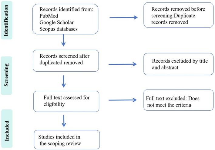
Relevant literature was retrieved from the PubMed, Google Scholar, and Scopus databases, covering the publication period from 2015 to 2025. This review included clinical trials and experimental studies conducted on animal models. Additionally, the reference lists of published systematic reviews and meta‐analyses were manually screened to identify additional eligible original studies that were not captured in the initial database searches. The search strategy incorporated the following keywords: "Alzheimer's disease", "Periodontitis", "Oral‐brain axis", "Neuroinflammation", "Aging", and "Bone remodeling". Boolean operators ("AND" and "OR") were strategically applied to optimize the search strategy, ensuring comprehensive retrieval of studies relevant to the research topic (Figure 1).

PRISMA flowchart.
Inclusion Criteria
Studies exploring the link between periodontitis and Alzheimer's disease (AD), encompassing both direct and indirect mechanistic pathways, were considered for inclusion. Articles focusing on the elderly demographic were prioritized, considering that aging constitutes a major shared risk factor for both conditions. Research that offers clinical, microbiological, or epidemiological data pertinent to the association between these two disorders was included.
Exclusion Criteria
Studies were excluded on the following grounds: failure to meet the predefined inclusion criteria, insufficient methodological rigor, unclear description of research methods, inadequate sample sizes, or poor reproducibility.
Overview of Alzheimer's Disease and Periodontitis
Alzheimer's Disease: Aging‐Related Hallmarks
AD is characterized by cognitive decline linked to hippocampal and cortical neuronal loss, with pathogenesis centered on three interconnected pillars: β‐amyloid (Aβ) aggregation, tau pathology, and chronic neuroinflammation [12]. Aβ peptides (predominantly Aβ40/42) are generated via aberrant proteolysis of amyloid precursor protein (APP) by β/γ‐secretases [13]; the highly hydrophobic Aβ42 readily forms extracellular plaques that impair synaptic plasticity critical for memory [14]. Tau, a microtubule‐stabilizing protein [15], undergoes hyperphosphorylation due to dysregulated kinases (GSK‐3β, CDK5) and reduced phosphatase (PP2A) activity, losing microtubule‐binding capacity and aggregating into intracellular neurofibrillary tangles (NFTs) with prion‐like pathological propagation [16]. Phosphorylated tau can act as a "prion‐like" protein, templating the conversion of native tau into pathological conformations, driving progressive neurodegeneration [17]. Chronic neuroinflammation, driven by dysfunctional microglia and reactive astrocytes, exacerbates neurodegeneration: activated microglia release pro‐inflammatory cytokines (IL‐1β, TNF‐α) and ROS, while reactive astrocytes form gliosis secreting context‐dependent neurotoxic or neuroprotective factors [18]. Though initially protective, sustained neuroinflammation in aging creates a pro‐degenerative microenvironment that amplifies Aβ/tau pathology. Together, these intertwined pathways—dysregulated proteolysis, tau misfolding, and chronic neuroinflammation—form a degenerative loop, exacerbated by aging‐related microglial dysfunction and blood–brain barrier (BBB) impairment, driving progressive synaptic damage and neuronal loss in AD.
Periodontitis: A Chronic Inflammatory Oral Disease
Periodontitis is a prototypical chronic inflammatory oral disease characterized by progressive destruction of periodontal tissues—including the gingiva, periodontal ligament, alveolar bone, and cementum—ultimately leading to tooth loss [19]. Affecting over 10% of the global population, it is marked by deep periodontal pocket formation, gingival inflammation, and irreversible loss of supportive structures. Its pathogenesis involves a complex interplay of microbial colonization, immune hyperactivation, and tissue‐destructive pathways.
Periodontitis initiates with the accumulation of dental plaque biofilm, a structured community of microorganisms [20]. Early colonizers (e.g., Streptococcus sanguinis, Actinomyces) adhere to the tooth surface, forming a glycoprotein pellicle. As the biofilm matures under anaerobic conditions, it transitions into a pathogenic consortium dominated by Gram‐negative anaerobes, including Porphyromonas gingivalis, Tannerella forsythia, and Treponema denticola (the "red complex"). These pathogens secrete virulence factors—such as gingipains produced by Porphyromonas gingivalis—that degrade host immunoglobulins, cytokines, and epithelial tight junctions, thereby impairing the integrity of mucosal barriers. The biofilm matrix protects bacteria from immune clearance and antimicrobial therapies, while quorum sensing coordinates the expression of virulence genes, enhancing pathogenicity.
The host response to pathogenic biofilms transitions from protective to destructive, driven by excessive inflammation and failed resolution. Innate immune receptors (TLR‐2/TLR‐4) recognize microbial components like LPS and peptidoglycan, activating the NF‐κB pathway and inducing pro‐inflammatory cytokines (IL‐1β, TNF‐α, IL‐6) that recruit neutrophils and macrophages to the periodontal pocket [21]. While neutrophils attempt to clear bacteria via myeloperoxidase and elastase, these enzymes also degrade collagen in the periodontal ligament, causing collateral tissue damage [22]. Concurrently, regulatory T cells (Tregs) are suppressed: Porphyromonas gingivalis degrades the Treg transcription factor Foxp3, reducing anti‐inflammatory signals (IL‐10, TGF‐β) [23]. This immune imbalance—marked by unchecked pro‐inflammatory responses and insufficient resolution—fuels persistent tissue destruction, despite ineffective antibody‐mediated pathogen clearance due to bacterial evasion mechanisms like intracellular persistence.
Periodontitis arises from a failure to resolve the conflict between pathogenic biofilms and dysregulated host immunity, leading to uncontrolled inflammation and tissue destruction. These factors create a self‐perpetuating cycle: pathogens induce inflammation, which damages tissues and creates a niche for further microbial invasion, while host aging and comorbidities erode protective responses. Additionally, systemic conditions like diabetes exacerbate periodontitis via advanced glycation end products (AGEs) activating RAGE receptors [24], while smoking inhibits neutrophil function and collagen synthesis [25]. This suggests a strong mechanistic linkage between periodontitis development and systemic diseases.
Epidemiological and Clinical Evidence of the Bidirectional Relationship
Epidemiological studies have established a significant dose‐dependent association between periodontitis and AD, with mounting evidence from longitudinal cohorts, case–control studies, and meta‐analyses [26, 27, 28, 29]. Individuals with periodontitis exhibit an increased risk of developing AD compared to healthy counterparts [30, 31, 32, 33]. A retrospective cohort study of 262,349 participants found chronic periodontitis (CP) was associated with a 5% higher risk of AD (aHR = 1.05, 95% CI = 1.00–1.11), highlighting CP as a potential modifiable risk factor for dementia independent of lifestyle behaviors [34]. The conclusion was also confirmed by a prospective cohort study of 90 ad patients, with periodontitis patients exhibiting accelerated cognitive decline [35]. Additionally, a quasi‐experimental study involving 177 periodontally treated patients and 409 untreated subjects found that periodontal treatment was associated with a favorable effect on AD‐related brain atrophy (adjusted effect: –0.41, 95% CI: −0.70 to −0.12), suggesting a potential role in mitigating preclinical AD [36]. These findings collectively highlight periodontitis as a modifiable risk factor with graded effects on AD susceptibility. However, a few inconsistent results have also been reported [36]. It has been hypothesized that these discrepancies are likely attributable to confounding variables, including variability in participant age, follow‐up duration, and baseline oral hygiene status. Prospective, standardized cohort studies with rigorous control for such confounders and uniform outcome measures are therefore warranted to resolve these inconsistencies and validate the proposed links.
Current research on the relationship between AD and periodontitis has primarily centered on the role of periodontitis in promoting the onset and progression of AD [6]. However, cross‐sectional data show that AD patients have higher bleeding on probing (BOP%) and greater plaque index (PLI) than age‐matched controls, reflecting a bidirectional association, whereby AD may also accelerate periodontal disease progression [37]. While the prevailing hypothesis attributes severe periodontal tissue destruction in AD patients to deteriorated oral hygiene maintenance due to cognitive dysfunction, recent clinical observations highlight potential synergistic interactions between neurodegeneration and periodontal inflammation [28]. These findings establish AD as a significant modifier of periodontitis progression, with epidemiological evidence linking cognitive impairment to accelerated tissue destruction [38]. Similarly, further epidemiological studies are still needed in the future.
Mechanistic Basis of the Bidirectional Association
Periodontitis to: Pathogen‐Mediated Neuroinflammation and Neuronal Damage AD
Periodontitis, a chronic bacterial inflammatory disease, involves periodontal pathogens whose pathogenic impacts extend well beyond localized tissue destruction. These microorganisms can translocate systemically via hematogenous, lymphatic, or neural pathways, enabling them to influence distant organ systems and contribute to the pathogenesis of extraporal diseases—processes collectively termed microbial translocation. Growing evidence indicates that Porphyromonas gingivalis and Treponema denticola—key pathogens of the periodontal "red complex"—can translocate to the brain, penetrating the BBB to induce neuroinflammation, synaptic dysfunction, and pathological cascades that contribute to cognitive impairment and AD [39, 40].
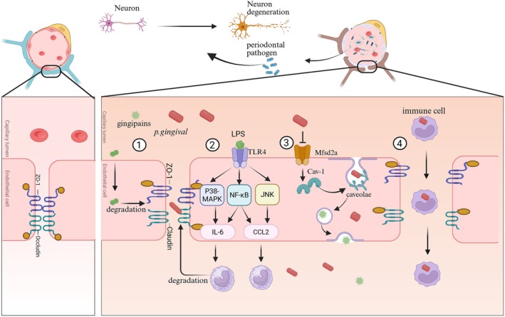
Multiple routes are exploited by periodontal pathogens to invade the brain by crossing BBB. One primary route is hematogenous spread, whereby periodontal pathogens gain access to the circulatory system via compromised gingival capillaries, especially during episodes of bleeding or biofilm perturbation. Once in circulation, certain periodontal pathogens are capable of directly breaching the BBB to invade the brain parenchyma [9, 41]. BBB, composed of brain microvascular endothelial cells (BMECs) with tight junctions and supported by astrocytic end‐feet, pericytes, and a basement membrane, serves as a critical protective interface between the circulatory system and central nervous system (CNS). Recent evidence demonstrates that Porphyromonas gingivalis enables direct promotion of the invasion of pathogens into the brain by increasing major facilitator superfamily domain containing 2a/Caveolin‐1 (Mfsd2a/Cav‐1)‐mediated transcytosis in BMECs [42]. Additionally, Porphyromonas gingivalis exploits virulence factors like lipopolysaccharides (LPS) to increase BBB permeability and allow bacterial extravasation into the brain by directly degrading tight junction proteins (e.g., ZO‐1 and occluding) [43]. The virulence factors can also enter the cerebral parenchyma and initiate the inflammatory response to compromise BBB integrity [44]. In addition to direct invasion, periodontal pathogens also coerce immune cells to cross the blood–brain barrier; extracellular vesicle (EV) membrane fusion plays a critical role in cargo delivery, enabling the transfer of microbial virulence factors [45]. Besides compromising BBB integrity, pathogens, shielded within EVs, can directly traverse the BBB via vesicle‐mediated immune evasion mechanisms, triggering the accumulation of Aβ, a hallmark of AD pathogenesis [46]. These results demonstrate that hematogenous spread is the primary route through which pathogens invade the brain. Beyond hematogenous mechanisms, trigeminal nerve retrograde transport serves as an alternative route. Xiaoyang Ma et al. found fluorescein‐labeled Porphyromonas gingivalis extracellular vesicles in trigeminal ganglia and hippocampus following gingival exposure, indicating the trans‐nerve migration potential of pathogens to the brain [47]. Recent investigations underscore the intestinal pathway as a critical route for periodontal pathogens to influence AD progression. A seminal study by a Nanjing University research group demonstrated that oral microbiota derived from periodontitis patients, when administered via gavage to healthy mice, induced significant dysregulation of intestinal microbial homeostasis and perturbed intestinal immune balance [48]. These findings establish the oral‐intestinal axis as a novel transmission mechanism, operating independently of the bloodstream, by which periodontal pathogens may modulate neuroinflammatory and neurodegenerative processes associated with AD pathogenesis (Figure 2).
Upon invasion of the brain, periodontal pathogens initiate a dual‐pronged pathogenic process—neuroinflammation and direct neuronal damage—that collectively drive cognitive impairment. Neuroinflammation is primarily triggered by the activation of innate immune cells in the CNS through pathogen‐associated molecular patterns (PAMPs) [49]. Virulence factors such as LPS and gingipains from Porphyromonas gingivalis bind directly to Toll‐like receptor 4 (TLR4) and protease‐activated receptors (PARs) on microglia, activating the nuclear factor kappa–light–chain–enhancer of activated B cells (NF–κB) and mitogen‐activated protein kinase (MAPK) signaling pathways [50]. This activation elicits a persistent release of pro‐inflammatory cytokines, including IL‐1β and TNF‐α [50]. Subsequently, astrocytes are secondarily activated within the inflammatory microenvironment; they secrete chemokines (e.g., CXCL10), exacerbating immune cell infiltration and establishing an inflammatory cascade [51]. Chronic neuroinflammation disrupts synaptic plasticity and neurotransmitter transmission, and it further upregulates inducible nitric oxide synthase (iNOS), generating excessive nitric oxide (NO). The resultant oxidative stress and additional BBB damage create a permissive environment for pathogen dissemination and neurodegeneration. As a core pathogen in chronic periodontitis, Tannerella forsythia also plays a critical role in the progression of AD [52]. The key surface virulence molecules of this bacterium include BspA protein (a leucine‐rich repeat adhesion protein) and capsular polysaccharides [53]. Among them, the BspA protein can activate the inflammatory response of microglia and astrocytes by binding to TLR2 on the surface of host cells [54, 55]. Notably, accumulating evidence indicates that Tannerella forsythia acts synergistically with Porphyromonas gingivalis in pathological processes. In particular, cell extracts from Tannerella forsythia can stimulate the growth of Porphyromonas gingivalis [56]. Considering that Porphyromonas gingivalis has been implicated in promoting the progression of AD, it is plausible that Porphyromonas gingivalis and Tannerella forsythia exert a synergistic effect, which may in turn exacerbate AD‐associated pathogenesis. Moreover, it has been documented that both Fusobacterium nucleatum and Aggregatibacter actinomycetemcomitans cooperate synergistically with Porphyromonas gingivalis to drive the progression of neuroinflammation. The three periodontal pathogenic bacterial species exhibit a prominent metabolic complementarity, which serves as a critical mechanistic basis for their synergistic pathogenicity. Specifically, Porphyromonas gingivalis is auxotrophic for exogenous heme and vitamin B12 for proliferation [57]. In contrast, Fusobacterium nucleatum can efficiently sequester heme from the oral microenvironment via the heme‐binding protein (HbpA) localized on its outer membrane, while Aggregatibacter actinomycetemcomitans possesses the biosynthetic machinery to endogenously produce vitamin B12 [58, 59]. Through a well‐orchestrated "metabolic sharing" mechanism, Fusobacterium nucleatum and Aggregatibacter actinomycetemcomitans respectively supply Porphyromonas gingivalis with the indispensable heme and vitamin B12, thereby directly fulfilling Porphyromonas gingivalis's nutritional requirements. This interspecies metabolic collaboration not only facilitates the robust proliferation of Porphyromonas gingivalis but also upregulates the transcriptional expression of Porphyromonas gingivalis's virulence‐associated genes—including kgp and rgp, which encode the cysteine proteases gingipains (lysine‐specific gingipain and arginine‐specific gingipain, respectively)—leading to a marked enhancement of Porphyromonas gingivalis's pathogenic potential. Upon infiltration into the CNS, Fusobacterium nucleatum, Aggregatibacter actinomycetemcomitans, and Porphyromonas gingivalis further synergize to trigger robust inflammatory responses. These pathogens release a repertoire of virulence factors, such as lipopolysaccharides, fimbriae, and toxins, which act in concert to activate two major CNS‐resident cell populations: microglia and astrocytes [60]. This coordinated activation elicits multi‐dimensional inflammatory signaling cascades, including the sustained activation of the TLR‐NF‐κB pathway, the assembly and activation of the NOD‐like receptor family pyrin domain‐containing 3 (NLRP3) inflammasome, and the initiation of the pyroptotic pathway. These inflammatory signaling events exhibit a mutually reinforcing and progressive amplification effect, ultimately culminating in a prominent "inflammatory amplification loop." This loop not only exacerbates the local inflammatory milieu within the CNS but also sustains the progression of neuroinflammation, thereby contributing to the pathogenesis of neuroinflammatory and neurodegenerative disorders (Table 1).
Direct neuronal damage by periodontal pathogens relies on their specialized virulence mechanisms [46]. Gingipains (RgpA and Kgp) secreted by Porphyromonas gingivalis, as cysteine proteases, selectively cleave neuronal surface receptors, extracellular matrix proteins, and key signaling molecules. Studies have demonstrated that gingipains degrade neural cell adhesion molecule (NCAM) and nerve growth factor (NGF) receptor TrkA, thereby disrupting inter‐neuronal connections and inhibiting axonal growth [47]. Additionally, gingipains process the β‐amyloid precursor protein into neurotoxic Aβ fragments and promote abnormal phosphorylation of tau protein, accelerating the formation of neurofibrillary tangles [66]. Anaerobic pathogens like Treponema denticola directly damage neuronal membranes through outer‐membrane proteins and hemolysins, inducing mitochondrial dysfunction and apoptosis. Recent research has revealed that EVs of periodontal pathogens can traverse the BBB, carrying virulence factors [45]. Through membrane fusion, EVs deliver gingipains and LPS directly into the neuronal cytoplasm, triggering caspase‐3‐dependent apoptosis and endoplasmic reticulum stress [61]. In addition, certain periodontal pathogenic bacteria can secrete specific virulence factors that directly target kinases associated with tau protein phosphorylation in neurons, triggering their abnormal activation and thereby accelerating the process of tau protein phosphorylation. Among these bacteria, Porphyromonas gingivalis has the most well‐defined role and the most sufficient research evidence [61]. The gingipains secreted by this bacterium not only cleave the APP to promote the production of Aβ, but also directly act on the key regulatory kinases of tau protein phosphorylation, namely p38 mitogen‐activated protein kinase and glycogen synthase kinase‐3β (GSK‐3β) [62]. Researchers found that Porphyromonas gingivalis could significantly upregulate the phosphorylation level of p38 MAPK [67]. The activated p38 MAPK could directly phosphorylate specific sites of the tau protein, which are exactly the main phosphorylation sites of the tau protein in NFTs in the brains of AD patients. And other periodontal pathogenic bacteria can also promote the process of tau protein hyperphosphorylation. Tannerella forsythia activates the MAPK inflammatory pathway by binding to immune cells' TLR2 through the BspA protein, thereby promoting the release of pro‐inflammatory factors. Fusobacterium nucleatum enhances the permeability of the blood–brain barrier, allowing peripheral inflammatory cells to infiltrate and activate, indirectly promote tau protein hyperphosphorylation [68]. Moreover, chronic neuroinflammation leads to abnormal function of astrocytes, reduces the supply of PP2A, forming a "inflammation‐insufficient phosphatase‐tau accumulation" vicious cycle, accelerating the formation of NFTs and neuronal death.
While Aβ and tau pathology have been the focus of periodontitis‐AD research, the involvement of α‐synuclein—a presynaptic protein aggregated in Parkinson's disease (PD) and dementia with Lewy bodies (DLB)—remains largely unexplored. α‐synuclein aggregation is driven by neuroinflammation, oxidative stress, and microbial insults [69], all of which are hallmarks of periodontitis. However, direct evidence linking periodontal pathogens to α‐synuclein misfolding or propagation is lacking. Several open questions persist: (1) Do periodontal pathogens (e.g., P. gingivalis, T. forsythia) or their virulence factors induce α‐synuclein phosphorylation or oligomerization in neurons or glia? (2) Can periodontitis‐associated neuroinflammation promote the prion‐like spread of α‐synuclein aggregates in the brain? (3) Is there an overlap between periodontitis‐induced tau/Aβ pathology and α‐synuclein aggregation, potentially exacerbating mixed dementia phenotypes? Preliminary in vitro data suggest that LPS from P. gingivalis may upregulate α‐synuclein expression in neuronal cells, but in vivo validation and mechanistic studies are absent. Given the comorbidity of AD and synucleinopathies in aging populations, clarifying the role of α‐synuclein in the oral‐brain axis is essential to fully understand the spectrum of periodontitis‐associated neurodegeneration.
In summary, these multi‐dimensional pathogenic pathways that extend from local infections to neurodegenerative diseases provide crucial theoretical basis for the development of prevention and treatment strategies for AD and other cognitive disorders.

Multiple pathways in which periodontal pathogenic bacteria invade the blood–brain barrier. (1) Virulence factors secreted byare able to increase BBB permeability by directly degrading tight junction proteins (e.g., ZO‐1 and occludin). (2) The virulence factors, such as LPS, can induce brain microvascular endothelial cells to secrete pro‐inflammatory chemokines via the P38‐MAPK, NF‐κB, and JNK signaling pathways. This process promotes the recruitment and accumulation of inflammatory cells while compromising the integrity of the blood–brain barrier by disrupting the tight junctions of endothelial cells. (3)directly facilitates the invasion of pathogens into the brain by enhancing major facilitator superfamily domain containing 2a/Caveolin‐1 (Mfsd2a/Cav‐1)‐mediated transcytosis in brain microvascular endothelial cells. (4) Periodontal pathogens also induce immune cells to transmigrate across the blood–brain barrier. P. gingivalis P. gingivalis
| Author | Main results | Methods of use | Research significance |
|---|---|---|---|
| Ciccotosto GD. et al. (2024) [] [40] | Chronic oral inoculation of female mice withalone induced all seven examined AD‐related brain pathologies,alone caused specific ones, and their coinoculation led to limited pathologies with lower pathogenicity.Porphyromonas gingivalisTreponema denticola | Animals: Forty 12‐week‐old female C57BL/6 mice, randomly assigned to 4 equal groupsIntervention: Gingival margin inoculation at maxillary molars withorPorphyromonas gingivalisTreponema denticolaObservation: IHC of Brain Tissue | The host brain response elicited by oral coinoculation was less than that elicited by each bacterium, suggesting coinoculation was less pathogenic |
| Lei S. et al. (2023) [] [42] | increases the permeability of blood–brain barrier endothelial cells via Mfsd2a/Cav‐1‐mediated transcytosis by the binding of gingipains to caveolin‐1 and the inhibition of major facilitator superfamily domain containing 2a expression.Porphyromonas gingivalis | Animals: 8‐week‐old healthy SD rats were randomly divided into three groups: high‐intensity group; low‐intensity group and control groupIntervention: injected intravenously withor PBSP. gingivalisObservation: Transmission electron microscopy and Immunohistochemical observation | Mfsd2a/Cav‐1 mediated transcytosis is a key pathway governing BBB BMECs permeability induced by, which may contribute to/virulence factors entrance and the subsequent neurological impairmentsP. gingivalisP. gingivalis |
| Nonaka S. et al. (2022) [] [43] | Gingipains are delivered into human cerebral microvascular endothelial cells (hCMEC/D3) likely via outer membrane vesicles (OMVs), and directly degrade tight junction proteins Zonula occludens‐1 (ZO‐1) and occludin (both intracellularly and in vitro) to increase the permeability of the hCMEC/D3 cell monolayer. | Infection of hCMEC/D3 cells with WTP. gingivalis | Providing insight into the mechanism by whichcrosses the blood–brain barrier (BBB) and potentially contributes to Alzheimer's disease (AD)‐related cognitive declineP. gingivalis |
| Elashiry M. et al. (2024) [] [45] | Extracellular vesicles derived from the gingiva of mice and humans with periodontitis induced bycontainantigens (e.g., RGP, Mfa‐1) and proinflammatory cytokines (IL‐1β, IL‐6), can penetrate the blood–brain barrier (BBB) both in vitro (human 3D BBB model) and in vivo (uninfected recipient mice, colocalizing with hippocampal microglia), and promote BBB permeability.Porphyromonas gingivalisPg | Periodontitis model: C57B6 mice +oral gavage (6 weeks, control included)Porphyromonas gingivalisEXO prep/characterization: Mouse (gingiva/brain) + human (PD/healthy gingiva) EXOs; NTA/Western blot/TEMIn vivo BBB penetration: Labeled mouse PD/Con EXOs → uninfected WT mice; IVIS/confocal trackingIn vitro BBB assays: Human 3D model; TEER/FITC‐dextran for PD EXO‐induced integrity/permeabilityComponent detection: Pg antigens (RGP/Mfa‐1) + cytokines (IL‐1β/IL‐6) in EXOs | Representing the first demonstration that oral microbial‐induced EXO may contribute to Alzheimer's disease (AD) pathogenesis by crossing the BBB |
| Ma X. et al. (2023) [] [47] | Gingival exposure toor its extracellular vesicles induces periodontitis, memory impairment‐like behaviors, hippocampal inflammation, reduced neuroprotective/BBB‐related protein expression, and gut dysbiosis with colitis, likely via pEV translocation to the brain through the trigeminal nerve and LPS entry via the periodontal blood pathway.Porphyromonas gingivalis | Cognitive behaviors were measured in the Y‐maze and novel object recognition tasks. Biomarkers were measured using ELISA, qPCR, immunofluorescence assay, and pyrosequencing | PG‐derived extracellular vesiclesmay be a remarkable risk factor for dementia |
| Lu J. et al. (2022) [] [48] | Gavage of periodontitis‐related salivary microbiota for two months impairs cognitive function, increases β‐amyloid accumulation and neuroinflammation, and induces gut microbial dysbiosis, intestinal pro‐inflammatory responses, intestinal barrier impairment, and systemic inflammation. | Sample collection: Isolate salivary microbiota from periodontitis patients and healthy individuals Animal intervention: Gavage periodontitis‐related salivary microbiota to APPswe/PS1ΔE9 (PAP) transgenic mice for 2 months Detection: Assess cognitive function, cerebral β‐amyloid accumulation, neuroinflammation, gut microbial dysbiosis, intestinal pro‐inflammation, intestinal barrier impairment, and systemic inflammation | Periodontitis may exacerbate AD pathogenesis through the gut‐brain axis via swallowed salivary microbiota, providing a novel perspective on AD etiology and intervention |
| Gong T. et al. (2022) [] [61] | Oral gavage ofouter membrane vesicles (Pg OMVs, 4 mg/kg) for 8 weeks impairs memory/learning in 14‐month‐old mice, accumulates in hippocampus/cortex, reduces BBB tight junction proteins (ZO‐1, occludin, claudin‐5), activates astrocytes/microglia, and induces IL‐1β, tau Thr231 phosphorylation, and NLRP3 inflammasome activation (inhibited by MCC950 in vitro), with microglia‐conditioned media enhancing N2a neuron tau phosphorylation (attenuated by MCC950).Porphyromonas gingivalis | Animal: 14‐month‐old mice → oral gavage of Pg OMVs (4 mg/kg) or saline (q.o.d., 8 weeks) Behavioral tests: Open field, Morris water maze, Y‐maze In vivo: BBB permeability, hippocampal tight junction proteins (ZO‐1/occludin/claudin‐5), neuroinflammation, tau Thr231 phosphorylation, NLRP3 inflammasome; Pg OMVs localization (hippocampus/cortex) In vitro: BV2 + Pg OMVs (5 μg/mL) ± MCC950 (NLRP3 detection); N2a + microglia‐conditioned media ± MCC950 (tau phosphorylation assessment) | Pg OMVs trigger AD‐like pathologies via NLRP3 inflammasome |
| Jiang M. et al. (2021) [] [62] | Chronic systemic exposure tolipopolysaccharide (PgLPS) activates glycogen synthase kinase (GSK)‐3β in microglia and neurons, induces microglial tumor necrosis factor (TNF)‐α production, thereby triggering neuronal tau hyperphosphorylation, neuroinflammation, and learning and memory deficits in APPNL‐F/NL‐F mice without altering amyloid (A)β1‐42 expressionPorphyromonas gingivalis | In Vivo Model: 10‐month‐old AD model Intervention: PgLPS (1 mg/kg, i.p., daily × 3 weeks) Behavioral Test: Passive avoidance test (learning/memory) Techniques: IHC, Western blotting, ELISA 2 In Vitro Cell Models: MG6 microglia, N2a neurons Assays: TNF‐α: qPCR (mRNA), Western blotting (protein) | Inhibiting GSK3β activation may help delay the periodontitis‐promoted pathological progression of Alzheimer's disease |
| Wu B. et al. (2024) [] [63] | AD patients exhibit an increased osteoclastogenesis signature in blood (positively correlated with impaired peripheral Aβ clearance by immune cells), while long‐term pharmacological blockade of osteoclasts with Alendronate in APP23 transgenic AD mice improves peripheral monocyte Aβ‐degrading enzyme expression, reduces Aβ deposition, and mitigates memory decline | Targeted analysis of public whole blood transcriptomes from AD patients to identify molecular signatures and pathways linked to OC hyperactivation Using APP23 TG AD mice, long‐term Alendronate‐mediated OC inhibition to evaluate effects on AD pathology (Aβ deposition, Aβ‐degrading enzyme expression) and memory function | Osteoclasts promote AD development/progression possibly via modulating peripheral immunity and highlighting the potential of osteoporosis prevention in alleviating cognitive burden |
| Weng Y. et al. (2020) [] [64] | Triggering Receptor Expressed on Myeloid Cells 2 (Trem2) is significantly upregulated in the alveolar bones of patients with chronic periodontitis, and it amplifies reactive oxygen species (ROS) signals in osteoclasts through a Trem2/DAP12/Syk‐dependent signaling cascade; furthermore, soluble Aβ42 oligomers (Aβo) in the periodontitis microenvironment can directly bind to Trem2 to enhance this signal and osteoclastogenesis, while conditional knockout of Trem2 in osteoclasts inhibits alveolar bone resorption in mice with periodontitis | Clinical sample analysis: RNA‐seq was used to detect Trem2 expression in alveolar bones of chronic periodontitis patients Animal model study: A periodontitis mouse model was established; Trem2 was conditionally knocked out in osteoclasts, and alveolar bone resorption was compared between knockout and control groups Molecular mechanism exploration: Molecular biology techniques verified the Trem2/DAP12/Syk cascade in osteoclast ROS signal amplification, and explored the interaction between Aβo and Trem2 and its regulatory effect on osteoclastogenesis | Trem2 is a potential target for the prevention and treatment of bone destruction in periodontitis and AD |
| Catalina Arévalo‐Caro et al. (2025) [] [65] | This study investigates the potential link between periodontal disease (PD) and Alzheimer's disease (AD) through the apolipoprotein E gene ε4 allele (APOE4)—a primary genetic risk factor for AD associated with chronic inflammatory conditions like PD, which potentiates AD development and progression | The Joanna Briggs Institute methodology and PRISMA guidelines were followed. The search included articles published in PubMed and Embase, focusing on human studies, and excluding case series, in vitro studies, reviews, and animal studies | APOE4 may link PD and AD through shared genetic variants, inflammatory pathways, and dyslipidemia |
to Periodontitis: Neurodegeneration‐Mediated Disruption of Bone Homeostasis AD
AD‐related neurodegeneration impairs oral health through autonomic and behavioral mechanisms, which is regarded as the primary cause of periodontal destruction in AD patients. However, several studies have unveiled the inherent link between AD and periodontitis, suggesting that beyond oral hygiene practices, AD itself influences the progression of periodontitis [70]. Alveolar bone loss represents a key clinical manifestation of periodontitis, with bone homeostasis imbalance serving as a critical pathological basis for bone mass loss. Existing literature demonstrates that AD patients exhibit disruptions in bone homeostasis, further corroborating the role of AD in driving periodontal disease progression [71].
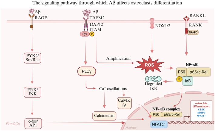
As a central pathologic hallmark of AD, Aβ in the brain can be excreted to peripheral organs and deposited in skeletal system, where it disrupts bone homeostasis—a process increasingly linked to aging‐related osteopenia. While direct evidence for Aβ exacerbating periodontitis progression remains inconclusive, a robust body of literature establishes its role in dysregulating bone remodeling and inducing bone mass loss, with mechanistic insights deeply rooted in aging‐associated cellular dysfunction. Aβ exerts age‐dependent effects on osteoclastogenesis, a key driver of bone resorption in aging populations [63]. Aβ promotes osteoclast differentiation and activation through multiple pathways: (1) engagement of the Receptor for Advanced Glycation Endproducts (RAGE), a receptor critically involved in age‐related inflammation, amplifies osteoclast‐mediated bone resorption [72]; (2) synergistic activation of NF‐κB, ERK, and calcium oscillation signaling enhances RANKL‐induced osteoclast activation, a pathway often dysregulated in senescent bone microenvironments [73, 74]; (3) interaction with Triggering Receptor Expressed on Myeloid Cells 2 (TREM2) initiates a reactive oxygen species (ROS)‐driven feedforward loop, accelerating osteoclast differentiation [64]; Notably, these pro‐resorptive effects are partially mitigated in aged individuals by upregulation of soluble RAGE (sRAGE) and osteoprotegerin (OPG), though this compensation coincides with impaired bone remodeling and increased fracture susceptibility—hallmarks of aging [72]. Concurrently, Aβ undermines osteoblast function and mesenchymal stem cell (MSC) fate commitment, critical processes for bone formation and renewal. Osteoblast‐specific APP overexpression in TgAPPswe‐OCN mice recapitulates trabecular bone loss [75], while young Tg2576 mice exhibit adipogenic skewing of MSCs, highlighting Aβ's dual role in suppressing osteogenesis and promoting marrow adiposity—phenotypes strongly associated with skeletal aging. Oxidative stress and mitochondrial dysfunction emerge as unifying mechanisms linking Aβ to skeletal dysfunction. Antioxidant interventions (e.g., N‐acetyl‐L‐cysteine) rescue osteogenic potential in Tg2576‐derived MSCs and alleviate osteopenia in vivo. Our recent work also demonstrates that Aβ‐induced mitochondrial fission impairs type H blood vessels—a key regulator of bone vascular osteogenesis—in APP/PS1 mice [76]. These findings converge on Aβ‐induced imbalance of bone homeostasis as a potential central node in periodontal destruction in AD patients (Figure 3).
As a critical component of the brain‐bone axis governing skeletal physiology, the autonomic nervous system (ANS) has emerged as a key regulator of bone metabolism. Notably, sympathetic nerve hyperactivity has been documented in AD patients [77], establishing a potential neuro‐skeletal connection. Preclinical studies have demonstrated that chemical sympathectomy alleviates inflammation‐induced alveolar bone loss [78], implicating excessive sympathetic activation as a modifiable target for bone preservation. Mechanistically, noradrenergic terminals within the bone microenvironment release norepinephrine (NE), which binds to β₂‐adrenergic receptors (β₂ARs) on osteoblasts and osteocytes. This signaling cascade exerts dual effects: (1) suppressing osteoblastic bone formation; and (2) upregulating receptor activator of nuclear factor kappa‐B ligand (RANKL) expression, thereby promoting osteoclastogenesis and enhancing bone resorption [79, 80]. Additionally, osteoclast progenitors themselves express β₂ARs; however, there is still some controversy over its direct role in sympathetic stimulation directly accelerates their differentiation into mature bone‐resorbing cells [81]. Complementing sympathetic function, the parasympathetic nervous system (PNS) maintains skeletal balance via counter‐regulatory mechanisms. While direct evidence linking PNS dysfunction to periodontitis in AD remains lacking, AD patients exhibit characteristic parasympathetic impairments—including gastrointestinal dysmotility and dysregulated blood pressure control [82]. These systemic manifestations suggest a broader role for PNS insufficiency in mediating inflammatory bone loss, potentially through disrupted neuro‐immune crosstalk in periodontal tissues.
Additionally, a multifaceted disruption of the endocrine system represents a critical pathway through which AD impacts bone remodeling, with core mechanisms embedded in the bidirectional interplay between neurodegeneration and dysregulated endocrine networks. These interactions fundamentally disrupt the dynamic equilibrium between bone formation and resorption, driving pathological skewed remodeling. Hyperactivation of the hypothalamic–pituitary–adrenal (HPA) axis in AD leads to sustained hypercortisolism [83], which directly inhibits osteoblast proliferation and differentiation while reducing bone matrix synthesis and enhances osteoclast precursor recruitment and activity to promote bone resorption [84, 85]. Cortisol also disrupts vitamin D metabolism by inhibiting renal proximal tubule 1α‐hydroxylase activity, decreasing intestinal calcium absorption and indirectly exacerbating bone loss [86]. Sex hormone imbalance plays a critical role in bone remodeling disorders: reduced estrogen levels in postmenopausal female AD patients weaken its inhibitory effect on osteoclasts (by decreasing secretion of osteoprotegerin, an inhibitor of receptor activator of nuclear factor κB ligand [RANKL]) and reduce osteoblastic sensitivity to growth factors [87], while testosterone deficiency in male AD patients impairs bone formation by downregulating androgen receptor expression in osteoblasts and inhibiting IGF‐1 synthesis, potentially synergizing with cortisol to exacerbate muscle loss and reduce skeletal mechanical load stimulation. As a hallmark of AD, glycometabolic dysfunction ("type 3 diabetes") impairs osteoblastic insulin receptor signaling via insulin resistance, inhibiting glucose transport into osteoblasts and collagen synthesis, while reduced glucagon‐like peptide‐1 (GLP‐1) secretion not only exacerbates glycemic fluctuations but also indirectly promotes bone resorption by weakening inhibition of osteoclasts [88]. Hypothalamic–pituitary‐growth hormone axis dysfunction, leading to deficiencies in growth hormone (GH) and insulin‐like growth factor‐1 (IGF‐1), directly impairs osteoblast proliferation and differentiation; IGF‐1 insufficiency further reduces type I collagen synthesis and inhibits osteoclast apoptosis, disrupting bone remodeling bidirectionally [88]. Thyroid dysfunction (e.g., low T3 syndrome) slows bone turnover and affects mineralization by downregulating thyroid hormone receptor β expression in osteoblasts [89]. Additionally, circadian rhythm disruption of melatonin secretion in AD may increase bone marrow adipocyte differentiation (marrow adiposity inhibits osteogenesis) via the suprachiasmatic nucleus‐sympathetic nerve pathway, while melatonin deficiency may weaken its inhibitory effect on osteoclast activity [88]. These endocrine abnormalities collectively act through multiple synergistic pathways, ultimately leading to osteoclast‐dominated bone resorption exceeding osteoblast‐mediated bone formation, inducing osteoporosis, bone microarchitecture deterioration, and increased fracture risk, thus creating a vicious cycle between AD and bone metabolic disorders.
Although the specific mechanisms described above remain to be validated in periodontitis, the endocrine‐mediated pathways through which AD influences bone metabolism may underlie the accelerated progression of periodontitis in individuals with preclinical AD. Prospective translational studies are therefore warranted to investigate this hypothesis and clarify the bidirectional relationship between neurodegenerative processes and periodontal bone loss.

Multiple signaling pathways through which Aβ affects osteoclasts. Aβ drives osteoclast differentiation and activation via multiple mechanisms: (1) interaction with the Receptor for Advanced Glycation Endproducts (RAGE)—a key mediator of age‐related inflammation—promotes the expression of osteoclast differentiation genes. (2) binding to Triggering Receptor Expressed on Myeloid Cells 2 (TREM2) initiates a reactive oxygen species (ROS)‐dependent feedforward loop or calcium oscillation signaling cascades that accelerate osteoclast differentiation.
Age‐Related Factors Amplifying the Bidirectional Relationship
Periodontitis andboth fall under the category of age‐related diseases and aging primes a bidirectional exacerbation between periodontitis andthrough two interlinked processes: immunosenescence and the chronic low‐grade systemic inflammation characteristic of aging. These aging‐related shared mechanisms converge to impair clearance of oral pathogens and brain protein aggregates while fostering a pro‐inflammatory microenvironment, creating a vicious cycle that amplifies disease progression in both systems. AD AD
With age, naïve T cell pools shrink, while memory T cells accumulate in a senescent state, exhibiting reduced proliferation and skewed cytokine profiles [90]. In periodontitis, this compromises Th1/Th17‐mediated defense against Porphyromonas gingivalis, leading to impaired macrophage activation and 25% reduced phagocytosis of Porphyromonas gingivalis in aged gingival tissues [91]. Concurrently, in AD, senescent T cells fail to regulate microglial pro‐inflammatory responses, while B cells produce low‐affinity antibodies against Aβ, decreasing humoral clearance of Aβ oligomers [92]. Autopsies reveal reduced Aβ antibody titers in AD patients with periodontitis, correlating with higher plaque burden.
Aging also impairs neutrophil chemotaxis and phagocytic efficiency, delaying migration to periodontal pockets and reducing bacterial killing. This allows Porphyromonas gingivalis to persist and secrete virulence factors (e.g., gingipains), accelerating tissue destruction [93]. In the brain, aged neutrophils and monocytes exhibit defective clearance of Aβ aggregates: peripheral blood monocytes from AD patients show reduced phagocytosis of Aβ42 and increased release of IL‐1β, fostering neuroinflammation. Neutrophil extracellular traps (NETs) in aged individuals are hyperactivated but dysfunctional, releasing DNA citrullination products that cross‐react with tau, promoting tangle formation [94].
Brain‐resident microglia undergo senescence in aging, adopting a dystrophic phenotype with reduced phagocytic capacity and heightened secretion of SASP factors. This "primed" state amplifies neuroinflammation, while dendritic cells (DCs) in both oral and brain tissues exhibit reduced MHC‐II expression and increased IL‐10 secretion, promoting immune tolerance to pathogens and protein aggregates [95]. In the periodontium, senescent DCs fail to activate protective T cell responses, allowing biofilm persistence; in the brain, they exacerbate Aβ‐induced microglial activation, forming a feedback loop of dysfunctional immunity [96, 97].
Genetic factors are innate determinants of the pathogenesis of AD and periodontitis [98]. Certain genetic variants increase the susceptibility to both diseases by regulating shared pathways such as immune response, inflammatory reaction, and metabolic function, serving as a "genetic link" connecting the two. The apolipoprotein E (APOE) gene is currently recognized as a common high‐risk gene for AD and periodontitis, and its ε4 allele exerts bidirectional disease‐promoting effects through multiple mechanisms [65]. APOEε4 accelerates the progression of AD through three pathways: first, it reduces Aβ clearance efficiency (APOEε4 has weak binding ability to Aβ and cannot effectively mediate the transport of Aβ across the BBB or its phagocytosis by microglia); second, it promotes Aβ aggregation (APOEε4 can induce Aβ to form more toxic oligomers); third, it exacerbates neuroinflammation (APOEε4 can activate microglia to secrete more pro‐inflammatory factors) [99]. APOEε4 is also considered to be closely related to the progression of periodontal disease [100]. However, at present, there is a lack of corresponding basic research to verify this conclusion. We speculate that APOEε4 may influence the progression of periodontitis through the following aspects. First, it impairs the phagocytic capacity of neutrophils and macrophages against periodontal pathogens (APOEε4 affects the lipid metabolism of immune cells, reducing their antibacterial activity) [101]; second, it exacerbates periodontal inflammation (APOEε4 can increase the expression of TNF‐α and IL‐1β in periodontal tissues, amplifying the inflammatory response) [102]; third, it may reduce the repair capacity of periodontal tissues. In addition, triggering receptor expressed on myeloid cells 2 (TREM2) is a key regulatory factor for microglia and macrophages [103, 104]. Its genetic mutations can impair the ability of microglia to clear Aβ, increasing the risk of AD; at the same time, this mutation also reduces the bactericidal effect of macrophages on periodontal pathogens, leading to increased susceptibility to periodontitis and more severe disease conditions.
Lifestyle is a core acquired influencing factor for the pathogenesis of AD and periodontitis. Unhealthy living habits such as smoking and sleep disorders drive the progression of both diseases by long‐term affecting systemic metabolism, immune status, and oxidative stress levels, and there is a synergistic effect among these factors, forming a "multifactorial superimposed" risk effect [105]. Smoking is the strongest modifiable risk factor and can cause cross‐system damage. Harmful substances in tobacco, such as nicotine, tar, and heavy metals, can cross the BBB and directly damage neuronal cell membranes and DNA; at the same time, smoking activates the systemic oxidative stress response, promoting the production of reactive oxygen species (ROS) in the brain and accelerating Aβ aggregation and abnormal tau phosphorylation; in addition, smoking also reduces cerebral blood flow (especially in cognitive‐critical brain regions such as the hippocampus), exacerbating cerebral ischemia and hypoxia and impairing synaptic plasticity. Furthermore, smoking inhibits the proliferation of gingival fibroblasts and collagen synthesis, impairing the repair capacity of periodontal tissues; tobacco smoke reduces the concentration of antibacterial components such as lysozyme and mucin in saliva, increasing the colonization of pathogens such as Porphyromonas gingivalis and Tannerella forsythia; at the same time, smoking also reduces blood supply to periodontal tissues, lowering the chemotaxis and phagocytic efficiency of neutrophils, resulting in more severe periodontal inflammation and faster alveolar bone resorption [98]. In addition, sleep disorders (e.g., insomnia, obstructive sleep apnea) also act as bidirectional risk factors for AD and periodontitis by affecting the "clearance‐repair" process [106, 107].
Systemic chronic diseases such as diabetes, cardiovascular disease (CVD), and chronic obstructive pulmonary disease (COPD) accelerate the progression of both AD and periodontitis through shared pathways of "chronic inflammation‐immune dysregulation‐metabolic disorder," forming a cross‐system pathological chain of "systemic disease → local disease (periodontitis) → brain disease (AD)" [108]. In terms of promoting AD, cerebral insulin resistance in type 2 diabetes mellitus (T2DM) inhibits the activity of Aβ‐clearing enzymes, advanced glycation end products (AGEs) produced by hyperglycemia damage neurons and the BBB, and cerebral microangiopathy reduces blood supply to cognitive‐critical brain regions [109]; cerebral microangiopathy (sclerosis, embolism) in CVD causes cerebral ischemia and hypoxia, promoting Aβ deposition and tau protein tangles, systemic inflammation (e.g., elevated C‐reactive protein, IL‐6) activates microglia, and hypertension also damages the BBB [110]; long‐term hypoxemia in COPD damages cognitive brain regions such as the hippocampus, pro‐inflammatory factors (TNF‐α, IL‐8) released from the lungs spread to the brain through an "inflammatory cascade," and comorbid sleep apnea further exacerbates Aβ clearance disorders [111]. In terms of promoting periodontitis, all three diseases exert their effects by impairing the local defense and repair capacity of the periodontium, enhancing pathogen virulence, or exacerbating inflammatory responses. Hyperglycemia in T2DM provides "nutritional support" for periodontal pathogens (e.g., Porphyromonas gingivalis) and also damages neutrophil phagocytic function and increases the level of inflammatory factors [21]; impaired vascular endothelial function in CVD reduces blood supply to periodontal tissues, coagulation disorders (e.g., platelet activation) increase the risk of periodontal bleeding, and anticoagulant drugs (e.g., aspirin) taken by patients may also affect the effect of periodontal treatment; coughing and expectoration in COPD patients increase the colonization of oral pathogens (e.g., Klebsiella pneumoniae, Pseudomonas aeruginosa), long‐term use of glucocorticoids inhibits immune function [112], and hypoxemia also reduces blood supply to the periodontium, promoting the progression of periodontitis.
Therapeutic Implications and Interventional Strategies
Oral care for patients with AD must be adjusted according to the disease stages, with the core principle of adapting to changes in patients' cognitive and self‐care abilities, and reducing the risks of oral diseases and related complications (Table 2).
In the early stage, the focus is on prevention and the establishment of independent care habits [113]. Since patients still have relatively intact cognitive function, fluoride therapy should be implemented to prevent dental caries, and the intervals between oral examinations and scaling should be shortened. Simple and durable restorations are preferred to avoid complex treatments in later stages. Adaptive tools such as electric toothbrushes and water flossers should be provided, and strategies like color marking or the 5S methodology (Sort, Set in order, Shine, Standardize, Sustain) can be used to assist with memory [114]. Meanwhile, both patients and caregivers should receive education on proper cleaning techniques and dietary control (e.g., reducing sugar intake). Additionally, attention should be paid to maintaining masticatory function—dental prostheses (fixed or removable) should be provided promptly for tooth loss, and guidance on prosthesis cleaning should be given [115].
In the moderate stage, the key lies in managing resistant behaviors and maintaining oral health status. Patients often experience significant cognitive decline and behavioral resistance (e.g., refusing tooth brushing or dental treatment) [116]. During dental procedures, mouth props should be used to prevent biting, semi‐supine or supine positions to reduce aspiration risk, and appointment durations should be shortened (e.g., 15–30 min per session). Atraumatic restorative techniques (ART) are recommended for caries treatment. The "tell‐show‐do" communication method (explaining, demonstrating, and performing) should be adopted to reduce resistance, and caregivers should take the lead in daily oral care (e.g., assisting with brushing). Xerostomia should be closely monitored, and artificial saliva should be used when necessary [117]. For patients with dentures, caregivers should check for looseness or damage regularly to prevent aspiration.
In the late stage, palliative care and emergency management are prioritized. Patients with severe cognitive impairment (e.g., inability to recognize relatives or perform independent activities) require simplified cleaning, such as wiping the oral mucosa with saline‐soaked cotton swabs. Local antimicrobial gels (e.g., metronidazole gel) can be used for oral infections, and oral analgesics for severe toothache [118]. A multidisciplinary team (including geriatrics and respiratory departments) should assess the risk of aspiration pneumonia; in cases of severe oral bacterial accumulation, oral cleaning under general anesthesia may be considered [119]. Only life‐threatening oral issues (e.g., severe maxillofacial infections) require emergency intervention. Caregivers should be educated on identifying infection signs and basic first aid, while ensuring gentle care to respect patients' dignity.
Overall, oral care for AD patients requires stage‐specific adaptation, with continuous guidance for caregivers to maintain oral health and improve patients' quality of life.
| Stage | Core objectives | Specific care measures | Key considerations | Reference citations |
|---|---|---|---|---|
| Early stage | Prevention + Development of Independent Care Habits | Implement fluoride therapy and shorten intervals between oral examinations and scaling Prioritize simple and durable restorative treatment options Provide adaptive cleaning tools and use memory aids such as color coding or the 5S methodology Conduct health education for patients and caregivers Promptly provide fixed/removable dental prostheses for edentulism and offer guidance on prosthesis cleaning | Maintain patients' independent care capabilities and avoid over‐reliance on caregivers prematurely; reinforce memory through simple and understandable methods to ensure the sustainability of care measures | [,,] [113] [114] [115] |
| Moderate stage | Managing Resistant Behaviors + Maintaining Oral Health Status | Dental procedures: Use mouth props to prevent biting, adopt semi‐supine/supine positions to reduce aspiration risk, and shorten appointment durations Recommend Atraumatic Restorative Techniques (ART) for caries treatment Adopt the "tell‐show‐do" communication approach and have caregivers lead daily oral care Monitor xerostomia closely and use artificial saliva if necessary Regularly inspect dentures for looseness or damage | Focus on alleviating patients' resistant behaviors with gentle and efficient operations; strengthen caregivers' care capabilities to prevent complications such as aspiration and xerostomia | [,] [116] [117] |
| Late stage | Palliative Care + Emergency Management | Adopt simplified cleaning methods Use topical antimicrobial gels for oral infections and oral analgesics for severe toothache Establish a multidisciplinary team to assess aspiration pneumonia risk; consider oral cleaning under general anesthesia for severe bacterial accumulation; Perform emergency interventions only for life‐threatening oral issues Train caregivers in infection sign recognition and basic first aid skills | Prioritize patient dignity with gentle care operations; address life‐threatening emergencies as the top priority; provide comprehensive support to caregivers to reduce complication risks | [,] [118] [119] |
Challenges and Future Directions
Most studies exploring the periodontitis‐AD relationship are observational, establishing associations but not causal links. High‐quality longitudinal cohorts and randomized controlled trials (RCTs) are critical to disentangle causation, as current evidence cannot definitively prove that periodontal infection drives AD pathogenesis or vice versa. Additionally, pathogen‐specific mechanisms remain unclear: while Porphyromonas gingivalis is most studied, the distinct roles of other periodontal pathogens (e.g., Tannerella forsythia, Fusobacterium nucleatum) and their synergistic effects in AD pathology are underinvestigated. Heterogeneity in microbial composition and disease stages further complicates understanding of specific bacterial contributions, highlighting the need for mechanistic studies dissecting species‐specific impacts on neuroinflammation and amyloid/tau pathology.
Developing oral microbiota‐based biomarkers, such as salivary DNA of periodontal pathogens, offers non‐invasive potential for early AD screening. Advanced techniques like metagenomic sequencing and machine learning can identify pathogen‐host interaction patterns, with preclinical studies linking elevated salivary Porphyromonas gingivalis DNA to brain amyloid load. Concurrently, exploring dynamic oral‐brain axis mechanisms in aging—inclusive of microbial translocation, systemic inflammation, and barrier dysfunction—facilitates interdisciplinary interventions. Integrated strategies combining periodontal therapy (scaling, probiotics), cognitive training, and microbiota modulation aim to disrupt bidirectional inflammatory loops, with ongoing trials assessing their impact on cognitive decline and oral health outcomes in at‐risk populations.
Conclusion
The bidirectional relationship between AD and periodontitis is deeply rooted in shared pathological mechanisms, including microbial translocation, neuroinflammation, and disrupted bone homeostasis, with aging exacerbating this interplay via immunosenescence and inflammaging. Epidemiologically, periodontitis increases AD risk by 1.5–2‐fold, while AD patients exhibit a 35% higher periodontitis prevalence, forming a reciprocal inflammatory loop. Pathologically, oral pathogens drive Aβ/tau pathology via hematogenous/neural invasion, whereas AD impairs oral mucosal immunity and bone remodeling. Clinical evidence supports periodontal therapy as a modifiable strategy to reduce systemic inflammation, yet causal links and pathogen‐specific mechanisms remain unclear. Future research must prioritize longitudinal trials, mechanistic dissection of the oral‐brain axis, and interdisciplinary interventions integrating dental, neurological, and geriatric approaches to develop targeted preventive strategies for aging populations.
Author Contributions
W.Z., H.L. and M.L. wrote and revised the manuscript. Y.C. and Y.L. were involved in collecting data. W.Z. and S.L. were drawing the figures. All authors read and approved the final version of the manuscript.
Funding
This study was funded by the TaiShan Scholars of Shandong Province (No. tstp20221160) to Li M, the Construction Engineering Special Fund of "Taishan Young Scholars" of Shandong Province (No. tsqn202103177), the Natural Science Foundation of Shandong Province (No. ZR202210210042) to Liu H.
Ethics Statement
The authors have nothing to report.
Conflicts of Interest
The authors declare no conflicts of interest.