
What this is
- (OP) is a complex metabolic bone disease influenced by genetic and environmental factors.
- Over 200 million people globally suffer from OP, with a prevalence of 18.3%.
- This review discusses the molecular mechanisms of OP and evaluates various treatment options, including Western and Chinese herbal medicine.
Essence
- is characterized by increased bone resorption and decreased formation, leading to reduced bone mass and higher fracture risk. This review examines the molecular mechanisms of OP and compares therapeutic strategies, highlighting the need for effective treatments.
Key takeaways
- Over 200 million individuals globally are affected by , with a prevalence of 18.3%. This highlights the widespread impact of the disease on public health.
- Genetic factors contribute to 50%−80% of the variance in bone mass, indicating a strong hereditary component in risk.
- Current treatments include Western medicine and Chinese herbal remedies, each with distinct advantages and challenges, underscoring the need for personalized treatment approaches.
Caveats
- Many studies on Chinese herbal medicines are still in animal model stages, limiting their immediate clinical applicability.
- Research gaps exist in understanding the specific mechanisms of traditional Chinese medicine in treatment, necessitating further investigation.
Definitions
- Osteoporosis: A metabolic bone disease characterized by decreased bone mass and increased fracture risk.
- Bone remodeling: The process involving bone resorption and formation to maintain bone integrity.
Simplified
Introduction
Osteoporosis (OP) is a global health challenge that severely affects survival and quality of life. OP is a metabolic bone disease, and its occurrence and development mechanism are very complex and related to a variety of factors, such as genes, immune factors, drug factors, inflammation, and age. OP can destroy the normal physiological structure of bone, leading to a reduction in human bone mass and a decrease in bone strength and a substantial increase in the risk of fracture. Data show that worldwide, more than 200 million people worldwide suffer from OP; additionally, the overall prevalence is 18.3%. In terms of global geographical distribution, people in Europe have a relatively high probability of osteoporotic fractures, affecting up to 34.8% of this population. From the perspective of sex, women, especially postmenopausal women, have a greater probability of OP than men do. A survey study revealed that the prevalence of OP among postmenopausal women in Kenya is as high as 26.4% [1 –5]. From 2008 to 2018, the incidence of OP in the Korean population aged 50 years and older was 18.4 cases per 1,000 people per year [6]. Data from 2018 revealed that the prevalence rate of OP among people aged 50 years and older in China was 19.2% [7]. OP affects a large patient population worldwide and is characterized by a high prevalence rate.
OP can be divided into primary OP and secondary OP, of which primary OP is related mainly to aging factors. The most common clinical OP patients are postmenopausal women, and secondary OP stems from various causes, including endocrine diseases and drug effects [8]. Modern medical treatments for OP aim to improve bone metabolism, promote bone formation, and inhibit bone resorption. Drugs that inhibit bone resorption include estrogen, bisphosphonate, monoclonal antibodies, and calcitonin, and drugs that promote bone formation include parathyroid hormone and active vitamin D [9]. These drugs have achieved good efficacy in clinical practice, alleviating symptoms of discomfort and improving quality of life. However, these drugs also have many drawbacks, such as high cost, frequent adverse reactions, and certain carcinogenicity. In recent years, there have been numerous reports on clinical treatment with and related research on traditional Chinese medicine for treating OP. However, most studies on Chinese herbal medicines remain at the animal experiment stage, and there are many challenges with regard to translating such medicines into clinical use. At present, the prevention and treatment of OP remain a global challenge that urgently needs to be addressed.
In light of this background, This review first introduces the theoretical relationship between bone and OP and elaborates on the molecular mechanisms of OP from the aspects of genetic factors, environmental factors, signaling pathways, and bone remodeling. Next, the article summarizes the progress in the diagnosis of OP and comprehensively reviews the treatment regimens for OP. Treatment regimens include Western medicine treatment, Chinese herbal medicine treatment, nonpharmacological management and emerging therapeutic strategies. This review focuses on the molecular mechanisms and therapeutic progress of OP; it explores in depth the advantages and disadvantages of Western medicine and Chinese herbal medicine treatments, highlights the challenges that Chinese herbal medicine treatment for osteoporosis must overcome, and reveals the gap between emerging treatment methods and clinical applications, as well as potential directions for osteoporosis research, aiming to provide valuable references for future research on the prevention and treatment of osteoporosis.
The fundamentals of bone and osteoporosis
The skeletal system is composed of 206 bones and is critical within the human body. First, the skeleton supports the shape of the body, forming the trunk and limbs and supporting movement; it also protects the brain, heart, lungs and other important organs, and some studies have shown that the skeleton has detoxification and endocrine functions [10 –12]. Bones are very hard and metabolically active. Bone development begins as early as the beginning of pregnancy, and as the human body grows and develops, bone development continues until the age of 20 [13, 14]. Bone is composed mainly of periosteum, osseous tissue, endosteum and bone marrow. The outermost layer of every bone is the periosteum, which is a double-layered membrane. The outer fibrous layer is mainly responsible for supporting the rigid structure of bone. The inner cambium is rich in bone progenitor cells, which are important for the initial growth and development of bone and the repair of future damage. Osseous tissue is a mineralized tissue located between the periosteum and endosteum. Although osseous tissue does not contain bone marrow, the presence of mesenchymal stem cells in bone tissue is similar to that in bone marrow. Compared with the first two components, relatively few studies have investigated the endosteum, which originates from the periosteum. However, unlike the periosteum, the endosteum is very thin and plays an irreplaceable role in bone; it can not only promote bone repair but also control bone overgrowth and prevent the production of ectopic bone. Located in the deepest part of the bone, bone marrow is a key organ comprising hematopoietic stem cells and can be used for bone regeneration. At the cellular level, bone cells are composed of osteoblast lineages and osteoclast lineages. Osteoblasts are responsible for the continuous regeneration of bone and originate from mesenchymal stem cells. The main function of osteoclasts is the absorption of bone with aging, and osteoclasts are derived mainly from hematopoietic stem cells, such as macrophages and multinucleated giant cells. During the growth and development of the human body, bone formation and bone absorption are dynamic processes, and only a balance between the two can maintain the normal physiological structure and body function of the bone [15, 16]. If the balance between bone resorption and bone formation in the body is disrupted for various reasons, osteoclasts are activated, the absorption of bone significantly increases, the function of osteoblasts, i.e., new bone formation, is suppressed, bone mass decreases, bone metabolism regulation is disrupted, and bone density decreases, resulting in OP [17, 18].
Molecular pathogenesis of OP
Understanding underlying molecular mechanisms is a prerequisite for disease treatment. This review introduces the molecular mechanisms of OP from four aspects: genetic factors, environmental factors, signaling pathways, and bone remodeling.
Genetic factors
Genetic factors can affect bone mass and structure. Studies have shown that 50%−80% of the differences in bone mass among individuals are determined by genes. Using genome-wide association study technology, nearly 600 genes related to OP and fractures have been identified [19, 20]. Genetic factors include Deoxyribonucleic Acid/Ribonucleic Acid (DNA/RNA) methylation, histone modification, nucleosome localization, noncoding RNA, and chromatin configuration. DNA methylation can suppress important transcription factors, in turn reducing the expression of related genes and affecting the growth activity of many cells in bone. Recent research on DNA methylation and OP has focused mainly on changes in bone mineral density and risk factor markers of OP. However, relatively little research has investigated the mechanism of methylation in human osteoblasts at the whole-genome level, and therefore, more research is needed to provide new directions for the diagnosis and treatment of OP [21, 22]. Among noncoding RNAs(ncRNAs), microRNAs(miRNAs) regulate gene expression related to cell growth and apoptosis, and miRNAs are important for the maintenance of bone homeostasis. Serum miRNA expression increases significantly in OP fracture patients. miRNAs belong to the ncRNA family. Members of this family can recruit histone modifiers to regulate gene expression. On the one hand, miRNAs can degrade mRNA, and on the other hand, they can inhibit the translation of target genes, thus affecting the occurrence and development of osteoblasts and OP. Research has confirmed that when miR-218 activity is decreased, osteoblast activity is simultaneously inhibited, and bone mass further decreases. miR-188 and miR-218 are considered therapeutic targets for the treatment of OP [23, 24]. Another form of genetic influence is histone modification. In the presence of relevant factors, histones undergo posttranslational modification, which further affects gene transcription [25, 26]. In the field of bone metabolic diseases, histone acetylation and histone methylation are hotspots in research. Methyltransferases and demethylases play crucial roles in regulating gene expression in osteoblasts and osteoclasts [27, 28]. In-depth research on the genetic mechanisms can provide insight on the pathogenesis of OP and new ideas for the treatment of OP.

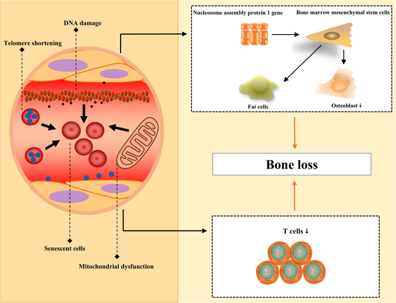
Aging of the body can cause OP. With increasing age, the number of senescent cells in the body gradually increases, and senile OP is a typical manifestation of senescence. Cell senescence is the result of many abnormal factors, including telomere shortening, DNA damage, and the physiological dysfunction of mitochondria. Under the influence of aging, the expression of the nucleosome assembly protein 1 gene is strongly affected, and the osteogenic differentiation ability of bone marrow mesenchymal stem cells is clearly suppressed. The aging of bone directly causes the abnormal transformation of bone marrow mesenchymal stem cells into fat cells, and T cells can also reduce the physiological function of cells during aging, resulting in bone loss. OP osteoporosis, DNA deoxyribonucleic acid
Environmental factors
OP results from the combined effects of genetic and environmental factors. Environmental factors include insufficient sunlight exposure, air pollution, dietary alterations, and unhealthy living habits, among others. Vitamin D plays an important role in the normal metabolism of bones. Ultraviolet B (UVB) light is an important environmental factor affecting the synthesis of vitamin D. Changes in season, latitude, and weather affect UVB exposure. Age, skin pigmentation, and duration of sun exposure are individual factors that influence vitamin D status (Wacker and Holick 2013). A cross-sectional study revealed that in the sun-deprived Sichuan Basin of China, the prevalence of vitamin D deficiency among adult women is very high in the winter; in sunny plateau areas, the 25(OH)D levels of young women with longer sun exposure are higher than those of their peers [40 –42]. Air pollution has been identified as a risk factor affecting bone density, and its adverse effects on bones are particularly significant in industrialized areas [43]. Data indicate that people who are exposed to long-term air pollution have a 15% increased risk of developing OP. Long-term exposure to fine particulate matter (PM) can activate receptor activator of nuclear factor-κB ligand 1 (RANKL). Moreover, long-term exposure to polluted air can promote the release of inflammatory factors. Furthermore, a polluted air environment can reduce the amount of UVB that reaches the Earth's surface, inhibit the formation of vitamin D, reduce bone formation, and affect the bone remodeling process [44]. Studies have shown that air pollution induces oxidative stress in osteocytes and that polluted fine particulate matter inhibits wingless-related integration site (Wnt) signaling activity in bones, thereby suppressing the bone formation process and reducing bone mass [45, 46]. Moreover, exposure to fine particulate matter is positively correlated with the serum levels of bone resorption markers. Air pollution can affect both bone resorption and bone formation, inhibit bone remodeling, and cause bone loss [47]. Smoking and alcohol consumption can also promote the occurrence of OP. Smokers have lower trabecular bone volume and average trabecular thickness, with reduced bone formation [48]. The chemical components in tobacco smoke have been indicated to cause bone mineral loss in animal models. In human experiments, smoking has been shown to be positively correlated with bone mineral loss, and the risk of fractures in smoking patients is significantly increased. Additionally, Parathyroid Hormone (PTH) levels in smokers are significantly reduced but return to normal levels after quitting smoking. Therefore, smoking can affect both bone resorption and bone formation [49, 50]. Compared with that among abstainers, the risk of osteoporosis among people who drink 0.5–1 cup of alcohol per day is 1.38 times greater, the risk of osteoporosis among people who drink 1–2 cups per day is 1.34 times greater, and the risk of osteoporosis among people who drink two cups or more per day is 1.63 times greater [51].
Signaling pathways

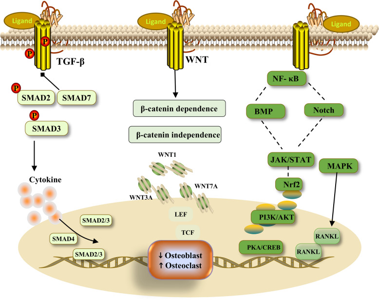
The abnormal activation of signaling pathways can affect bone metabolism. TGF-β can activate downstream SMAD signaling and interact with a variety of cytokines to affect bone metabolism. The Wnt signaling pathway can be divided into β-catenin-dependent and β-catenin-independent pathways. In the study of the human body, there are up to 19 Wnt ligands. If the activity of any Wnt signaling pathway in the body is impaired, normal bone metabolism is affected. The NF-κB and Notch signaling pathways inhibit osteoblast development and promote osteoclast differentiation. MAPK signaling mainly includes the ERK signaling, JNK signaling and p38 MAPK signaling pathways. Regulated by RANKL, MAPK promotes the differentiation of osteoclasts. In addition, other signaling pathways, such as the JAK/STAT, BMP and Nrf2 pathways, affect bone metabolism. TGF-β transforming growth factor-β, SMAD suppressor of mother against decapentaplegic, Wnt wingless-related integration site, NF-κB nuclear factor kappa-B, MAPK mitogen-activated protein kinase, ERK extracellular regulated protein kinases, JNK c-Jun n-terminal kinas, RANKL receptor activator of nuclear factor kappa-B ligand, JAK/STAT janus kinase/signal transducer and activator of transcription, BMP bone morphogenetic protein, Nrf2 nuclear factor erythroid 2-related factor 2,PI3K/AKT phosphoinositide 3-kinase/protein kinase B,PKA/CREB protein kinase A/cAMP response element binding protein,LEF lymphoid enhance factor,TCF t-cell factor
Bone remodeling process
The bone remodeling process is important for maintaining bone integrity and can be divided into four stages, namely, activation, resorption, reversal, and formation, that maintain bone stability. Under normal circumstances, osteoblasts and osteoclasts maintain a dynamic balance, thereby sustaining coordinated bone resorption and bone formation. In the OP state, bone resorption increases, bone formation decreases, the balance between osteoblasts and osteoclasts is disrupted, and bone mineral density decreases. Many factors affect the bone remodeling process, and complex molecular mechanisms interact with each other.
Oxidative stress

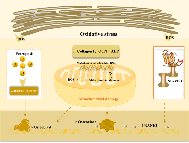
Oxidative stress plays an important role in regulating OP. Under oxidative stress, ROS increase abnormally and can significantly reduce the activity of Runx2 and osterix and affect the development of osteoblasts. Abnormal ROS accumulation can induce ferroptosis, further affecting osteoblasts. ROS can damage the normal function of mitochondria, and the loss of normal mitochondrial function affects neighboring mitochondria, leading to the release of a large number of ROS and inducing OP. Mutations in mitochondrial DNA are also associated with abnormal bone metabolism. Additionally, under oxidative stress, the activity of many antioxidant enzymes, such as collagen I, OCN and ALP, decreases, and osteogenic differentiation is affected. NF-κB signaling and RANKL activity are increased, and osteoclast differentiation is increased. OP osteoporosis, Runx2 runx family transcription factor 2, ROS reactive oxygen species, OCN osteocalcin, ALP alkaline phosphatase, NF-κB nuclear factor kappa-B, RANKL receptor activator of nuclear factor kappa-B ligand
The immune system and inflammation

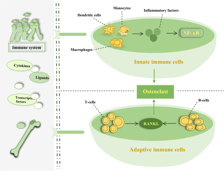
There is a strong link among the immune system, inflammation and OP. The skeletal system and the immune system have the same cytokines, transcription factors and ligands. Immune cells can be divided into innate and adaptive types. Innate immune cells include dendritic cells, monocytes, and macrophages; these cells promote the progression of inflammation, thereby increasing the function of osteoclasts and promoting bone loss. Representative inflammatory factors, including IL-6 and TNF-α, activate the NF-κB signaling pathway, causing inflammatory factor responses downstream of the signaling pathway and further stimulating the function of osteoclasts. Adaptive immune cells include T lymphocytes and B lymphocytes. In the absence of stimulation, T lymphocytes inhibit osteoclast function; if T lymphocytes are activated by RANKL, they play a role in strengthening osteoclasts. B cells can produce RANKL, which directly promotes bone loss. OP osteoporosis, NF-κB nuclear factor kappa-B, RANKL receptor activator of nuclear factor kappa-B ligand, IL-6 interleukin-6, TNF-α tumor necrosis factor-alpha
Iron metabolism
Iron is essential for maintaining normal physiological activities in the body; it is involved in cell growth and development and is also a key component of the oxidative stress system. The normal metabolism of iron in the body is complex and balanced and includes the production of iron, the utilization of iron by cells and tissues, the storage of surplus iron and the consumption of iron. Iron, which the body takes in through the diet, is first absorbed by the intestinal epithelium and then participates in various cycles for use by the body or is stored in ferritin. The iron transporter-sideromodulin regulatory axis regulates iron in the body to maintain an iron balance. If iron accumulates abnormally in the body, sideromodulin is activated, preventing iron from entering the circulatory systems in various ways [94, 95]. Iron can promote the production of free radicals, and ferrous iron can generate hydroxyl radicals, which are ROS. If the body is overloaded with iron and abnormal ROS accumulation occurs because of a variety of factors, the oxidative stress response is activated, resulting in the disruption of cytokines and many biological systems [96]. Abnormal iron metabolism, such as an abnormal increase in iron production or an abnormal decrease in iron excretion, can trigger oxidative stress and cause ferroptosis. Ferroptosis plays a regulatory role in a variety of diseases, such as malignant tumors, liver diseases, and cardiovascular diseases. Ferroptosis is associated with OP at the same time [97, 98]. First, excessive iron accumulation results in excess ROS production, activates oxidative stress, affects a variety of normal signaling pathways, and inhibits the function of osteoblasts; ferroptosis also significantly suppresses the function of bone marrow mesenchymal stem cells and further suppresses the formation of osteoblasts. Scholars have conducted in vitro experiments to assess the effects of ferroptosis on the mouse calvarial preosteoblastic cell line, subclone-E1(MC3T3-E1), and the results revealed that the osteogenic differentiation ability of MC3T3 cells under conditions of ferroptosis significantly decreased. In addition, iron metabolism affects osteoclasts. Iron accumulation leads to excessive ROS accumulation, which causes the abnormal activation of the MAPK and NF-κB signaling pathways, promotes the function of osteoclasts, and causes bone loss and OP [99, 100]. Iron metabolism is also involved in the ROS and oxidative stress systems. The connections among various systems, cells, and tissues in the body are complex and mysterious. Oxidative stress occurs mainly when the balance between oxidation and antioxidation is disrupted. Many inducing factors, including physiological and pathological factors, ultimately lead to an abnormal increase in ROS levels. An abnormal increase in ROS levels subsequently affects iron metabolism. In the case of abnormal iron metabolism, which is due mainly to iron overload, excessive ROS are produced, activating oxidative stress. Iron metabolism, oxidative stress, and ROS are closely connected. However, the role of ROS in iron metabolism and oxidative stress differs.
Gut microorganisms
The gastrointestinal tract has a large number and variety of microorganisms, and the intestinal microbiota has a symbiotic relationship with cells to maintain many physiological functions within the body. The gut microbiome includes bacteria, viruses, and fungi, which coexist harmoniously in the gut. The dysregulation of gut microbes is associated with a variety of diseases, such as diarrhea, obesity, and metabolic diseases. Once the gut microbiota is established, it remains relatively stable. As the body grows and develops, several factors affect the composition of gut microbes, such as diet, drug use, and inflammation [101 –103]. The intestinal microbiota not only affects the environment and physiological functions of the adjacent intestine but also extensively affects the physiological activities of bones, the liver, the brain and other organs. Inflammatory bowel disease, metabolic diseases, arthritis, rheumatoid arthritis, OP and other diseases are related to intestinal flora disorders [104, 105]. The gut microbiota plays important roles in bone loss and bone metabolism. Gut microbes can affect the skeletal system in a variety of ways. The skeletal system requires various hormones, such as parathyroid hormone, estrogen, and gastrointestinal hormones, to maintain its basic physiological activities. The production of these hormones is regulated by the intestinal microbiota, and metabolic disorders involving each hormone affect normal bone metabolism. For example, estrogen deficiency can cause an imbalance in the intestinal flora, which subsequently affects the skeletal system and causes bone loss, and estrogen supplementation can have a favorable effect by restoring the normal environment of the intestinal flora and inhibiting bone loss [106, 107]. An imbalance in intestinal microbes leads to increased intestinal permeability and damage to intestinal compaction-linked proteins, which further causes bacterial displacement and the spread of inflammation. In intestinal models, inflammation is significantly positively correlated with bone loss. Some bacteria among the intestinal microbiota can stimulate T helper 17 cell (Th17 cell) production, which can stimulate inflammatory cell Interleukin 17 (IL-17) secretion and RANKL production, further increasing osteoclast activity and bone absorption [108]. Short-chain fatty acids, mainly acetate, propionate and butyrate, are metabolites of intestinal microbial fermentation of dietary fiber. Short-chain fatty acids play important roles in maintaining normal intestinal permeability and the intestinal barrier, which can reduce the production of inflammatory factors and prevent bone loss. In addition, short-chain fatty acids directly affect osteoblasts, regulate osteoblast formation, promote bone formation, and inhibit osteoclast function. Short-chain fatty acids also promote the differentiation of bone progenitor cells by regulating the function of bone marrow stromal cells, further regulating bone metabolism, and maintaining normal bone density and bone health [106, 109].
Autophagy
Autophagy is a self-degrading process within cells, and its process is very conserved. Autophagy plays an important role in the maintenance of cell function and is directly involved in the development of cells and the maintenance of their renewal ability. There are three forms of autophagy, namely, macroautophagy, chaperone-mediated autophagy and microautophagy. Autophagy allows cells to continue growing even when there is a nutrient deficiency. Autophagy can regulate immunity and play a regulatory role in a variety of diseases, such as neurological diseases, cardiovascular diseases and musculoskeletal diseases [110]. Osteoblasts and osteoclasts maintain a normal autophagic state to maintain viability and balance bone metabolism. The disruption of the autophagic balance affects bone metabolism and causes OP. Reduced autophagy in osteoblasts reduces their lifespan. Osteocytes are terminally differentiated cells of the osteoblast lineage. Abnormal autophagy can lead to abnormal osteocyte function. Autophagy is involved in the regulation of bone metabolism by various growth hormones. Mitophagy can inhibit oxidative stress, regulate the functions of osteoblasts and osteoclasts, and maintain normal skeletal physiological activities [111]. In addition, multiple signaling pathways disrupt the differentiation balance between osteoblasts and osteoclasts by influencing the level of autophagy [112].
Other factors
Many other factors may affect the bone remodeling process, such as postmenopausal body status, excessive smoking, excessive alcohol consumption, glucocorticoid use, and the use of certain medications. The main cause of OP in postmenopausal women is estrogen deficiency. Glucocorticoids can increase the production of Peroxisome Proliferator-Activated Receptor Gamma 2 (PPAR-2), resulting in limited osteoblast growth, reduced bone formation, and bone loss. Compared with a control group, an experimental group receiving glucocorticoid treatment had significantly lower bone density. In addition to glucocorticoids, drugs such as proton pump inhibitors, chemotherapeutic agents, and antidepressants may affect bone remodeling, cause osteopenia, and induce OP [113].
In summary, OP is the result of the combined effects of environmental and genetic factors; its molecular mechanisms involve signaling pathways, oxidative stress, inflammation, intestinal microbiota, genes, autophagy, and other processes. The influencing factors are complex and can interact with each other, leading to increased bone resorption, decreased bone formation, imbalanced bone remodeling, and reduced bone mass.
Diagnosis of osteoporosis
The diagnosis of osteoporosis requires a combination of medical history, symptoms, and auxiliary examination results. Medical history includes personal history, family history, medication history, etc., for example, age, sex, medication use, and genetic disease history. Symptoms include bone pain, fractures, and limited mobility. Auxiliary examination results include laboratory tests and imaging examinations. Laboratory tests include routine blood tests, liver and kidney function tests, and electrolyte, alkaline phosphatase, 25-hydroxyvitamin D3, parathyroid hormone, and routine urine tests. Imaging examinations include X-ray, Computed Tomography (CT), Magnetic Resonance Imaging (MRI), nuclear medicine, and bone mineral density (BMD) [114]. X-ray and CT are common imaging methods for diagnosing OP. X-rays are more widely used. MRI can avoid ionizing radiation, but standards for the diagnosis of OP are lacking. Nuclear medicine examinations are highly important for the diagnosis of secondary OP. BMD measurement methods include dual-energy X-ray absorptiometry (DXA), quantitative computed tomography, and peripheral DXA. Among them, DXA is the most widely used. A Chinese expert consensus indicates that OP can be diagnosed when BMD measurements and clinical symptoms suggest OP; however, if imaging examinations indicate an insufficiency fracture, osteoporosis can be diagnosed [115]. The diagnostic criteria for osteoporosis established by the World Health Organizatio (WHO), which are based on DXA, are mainly for postmenopausal women, women who have undergone oophorectomy, and men over 50 years old [114].
In addition, the DXA trabecular bone score (TBS), spectral CT, dual-energy CT, MRI measurements of fat, and AI technology have provided new options for the diagnosis of OP. The above technologies have improved the ability to predict fracture risk. However, owing to equipment access and cost, more extensive research should be conducted on diagnostic applications for OP [116 –119].
Therapeutic interventions for osteoporosis
The treatment of OP is extensive and can be divided into Western medicine treatment, Chinese herbal medicine treatment, nonpharmacological management and emerging therapeutic strategies.
Therapeutic interventions involving Western medicine
There is no unified standard for the drug treatment of OP, thus requiring individualization in clinical practice. Drugs for treating OP can be divided into four main categories: antiresorptive drugs, anabolic drugs, dual-action drugs and other mechanism-based drugs. Currently approved drugs include bisphosphonates (alendronate, ibandronate, and zoledronic acid), parathyroid hormone analogs (teriparatide and abaloparatide), estrogen-related therapy (raloxifene-conjugated estrogens/bazedoxifene), RANK-ligand inhibitors (denosumab), sclerostin inhibitors (romosozumab), and calcitonin salmon [120]. Among them, parathyroid hormone analogs are anabolic drugs, sclerostin inhibitors are dual-action drugs, other drugs include antiresorptive drugs, and drugs with other mechanisms include active vitamin D and its analogs [121].
Bisphosphonates are traditional drugs for the treatment of OP. They can bind to bone minerals, inhibit the function of osteoclasts, and thus increase bone mass. Bisphosphonates are available in various formulations, including oral tablets, effervescent tablets, intravenous preparations, etc. They are inexpensive and are very commonly used as first-line drugs for the clinical treatment of OP. However, bisphosphonates cause adverse reactions, such as acute-phase injection reactions and gastrointestinal discomfort, and there is also a risk of osteonecrosis of the jaw. Denosumab is a biological agent and a human monoclonal antibody; it can inhibit RANKL activity and further suppress the formation of osteoclasts [122]. Raloxifene is an estrogen agonist that can reduce bone resorption and increase bone mineral density. Women with conditions such as venous thromboembolism, who are breastfeeding, and who are pregnant should avoid using raloxifene [123]. Calcitonin is a synthetic polypeptide hormone that can inhibit the function of osteoclasts; currently, it is not a first-line treatment for osteoporosis [124]. Relatively speaking, anabolic drugs and dual-action drugs are emerging as OP treatment options. Anabolic drugs include teriparatide and abaloparatide, both of which are parathyroid hormone analogs; they can stimulate osteoblasts, simultaneously stimulating Insulin-like Growth Factor-I (IGF-I)synthesis and inhibiting sclerostin, thus promoting bone formation [125]. Teriparatide was first applied in clinical practice and was the first approved osteogenic agent [126]. Abaloparatide was approved for clinical use by the U.S. Food and Drug Administration (FDA) in 2017; it binds to the same receptor as teriparatide does [127]. Dual-action drugs can promote bone synthesis while inhibiting bone resorption. Sclerostin is a glycoprotein secreted by osteocytes that inhibits Wnt signaling, leading to weakened osteoblast function. In addition, sclerostin enhances the activity of RANKL, resulting in increased osteoclast activity [128]. Currently, there are three anti-sclerostin monoclonal antibodies: blosozumab, setrusumab, and romosozumab. All of them have achieved good results in clinical trials. Romosozumab has been put into clinical use [129]. Research has shown that, compared with bisphosphonates, teriparatide and romosozumab lead to faster and greater increases in BMD, along with a substantial reduction in fracture risk [130, 131]. In terms of cardiovascular safety, real-world evidence has shown that both abaloparatide and teriparatide are safe [132]. However, compared with alendronate, romosozumab increases the risk of adverse cardiovascular events [133]. The European Medicines Agency has proposed that women who have had a myocardial infarction or stroke should avoid using romosozumab [134].
| Drug name | Target | Development stage | References |
|---|---|---|---|
| Alendronate sodium | FPPS | Marketed | [] [136] |
| Ibandronate sodium | FPPS | Marketed | [] [137] |
| Pamidronate sodium | FPPS | Marketed | [] [138] |
| Risedronate sodium | FPPS | Marketed | [] [130] |
| Zoledronic acid | FPPS | Marketed | [] [139] |
| Minodronic acid | FPPS | Marketed | [] [140] |
| Raloxifene | ER | Marketed | [] [141] |
| Denosumab | RANKL | Marketed | [] [142] |
| Teriparatide | PTHrP | Marketed | [,] [143] [144] |
| Abaloparatide | PTHrP | Marketed | [] [145] |
| Salmon Calcitonin | CGRP | Marketed | [] [146] |
| Eel Calcitonin | CGRP | Marketed | [] [147] |
| Ergocalciferol | VDR | Marketed | [] [148] |
| Cholecalciferol | VDR | Marketed | [] [149] |
| Calcifediol | VDR | Marketed | [] [150] |
| Calcitriol | VDR | Marketed | [] [151] |
| Alfacalcidol | VDR | Marketed | [] [152] |
| Eldecalcitol | VDR | Marketed | [] [153] |
| Romosozumab | SOST | Marketed | [] [154] |
| JMT103 | RANKL | Ph2(NCT05278338) | * |
| Setrusumab | SOST | Ph3(NCT05125809) | * |
| Blosozumab | SOST | Ph2(NCT02109042) | * |
| AMG 167 SHR-1222 | SOST SOST | Ph1(NCT01101048) Ph1(NCT04435158) | * * |
| Senolytics (Dasatinib + Querceti) | SASP | Ph2(NCT04313634) | * |
Therapeutic interventions with traditional Chinese medicine
| Therapeutic drugs | Research object | Mechanism | References |
|---|---|---|---|
| Andrographolide | Rats | ↓Senility | [] [156] |
| Dendrobium officinale | Senility mice | ↓Senility | [] [157] |
| Resveratrol | BMSCs | ↓Senility ↓ROS | [] [159] |
| Emodin | OP rats | ↓Inflammation | [] [161] |
| Eucommia ulmoides | OVX-OP rats | ↓Inflammation | [] [162] |
| Ginsenoside | DEX-OP rats | ↓Inflammation | [] [164] |
| Icariin | Mice and MC3T3-E1 cells | ↓NF-κB and RANKL | [] [167] |
| Quercetin | MC3T3-E1 cells | ↑Nrf2/HO-1 ↓ROS | [] [169] |
| Matrine | BMMSCs and C57BL/6-mice | ↓NF-κB and ↓AKT | [] [171] |
| Polydatin | OP mice | ↓MAPK | [] [172] |
| Artesunate | LPS-stimulated osteoclasts Mice | ↓TLR4/TRAF6 ↓PLCγ1-Ca | [] [173] |
| Crocin | Rats | ↓TNF-α and IL-6 ↓ROS | [] [176] |
| Cordycepin | BMSCs and OVX -mice | ↓ROS | [] [177] |
| Gastrodin | Iron overload mice Hippocampal neurons | ↑Nrf2/ARE ↓Ferroptosis | [] [178] |
| Curculigoside | MC3T3-E1 cells and mice | ↓ROS and ferroptosis | [] [179] |
| QinE pill | OVX-OP rats | ↑AKT/PI3K ↓Ferroptosis | [] [180] |
| Ginseng | BALB/c mice | ↑Intestinal flora ↓Intestinal permeability | [] [182] |
| Puerarin | OVX-OP rats | ↑Intestinal barrier ↑Short-chain fatty acid | [] [81] |
| Paeoniflorin | MC3T3-E1 cells C57BL/6-mice | ↑Autophagy | [] [187] |
| Cistanoside A | OB cells | ↑Autophagy | [] [188] |
| Tanshinone IIA | C57BL/6-mice BMMSCs | ↑Transcription factor NFATc1 | [] [185] |
| Puerarin | MC3T3-E1 cells | ↓TRPM3/miR-204 | [] [186] |
Nonpharmacological management
Nonpharmacological management for OP includes smoking cessation, limiting alcohol and caffeine intake, and exercise, among other strategies [121]. Exercise therapy is very practical; it can increase bone density and reduce the risk of fractures; however, the choice of exercise should be individualized [191]. Exercise methods include aerobic exercise, resistance exercise, and impact training. Aerobic exercise includes jogging, swimming, and Pilates. Traditional Chinese medicines such as Taijiquan and Baduanjin also include aerobic exercise. Resistance exercise includes weightlifting, push-ups, and impact training, such as skipping rope [192, 193]. Research has shown that for postmenopausal women with OP, regular light to moderate walking exercise can decrease physical pain, increase walking speed, enhance physical balance and increase daily activity ability [194]. Compared with high-intensity interval training or vitamin D alone, the combined treatment of high-intensity interval training and vitamin D supplements significantly increases bone mineral density in OP patients [195]. Another study revealed that exercise alleviates OP by regulating the secretion of the senescence-associated secretory phenotype [196]. Research has shown that ultraviolet therapy and extracorporeal shock wave therapy can increase bone mass in OP patients. Microwave therapy and medium-frequency or low-frequency pulses have good pain-relieving effects in OP patients [197].
Emerging therapeutic strategies
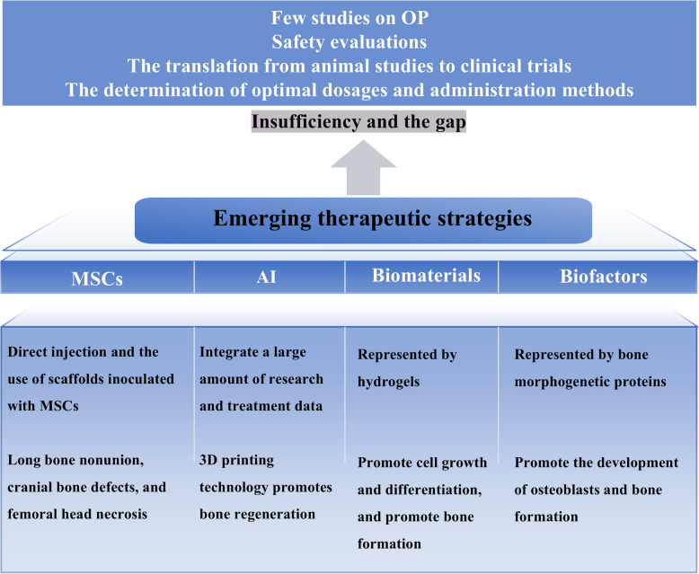
In summary, each of the four treatment methods has its own advantages and potential. Western medicine treatment is the most well developed and still occupies a dominant position. Some proprietary Chinese medicines have also been put into clinical practice and have achieved good clinical results. Nonpharmacological treatment is very practical and can be used as an auxiliary treatment method. Emerging treatment methods provide new ideas for the treatment of OP.

Emerging treatment options and their related shortcomings. There are four emerging treatment options: MSCs, AI, biomaterials, and biofactors. There is still a significant gap between emerging treatment options and clinical application, for example, few studies on OP, the translation from animal studies to clinical trials, the determination of optimal dosages and administration methods, and safety evaluations. MSCs marrow mesenchymal stem cells, AI artificial intelligence, OP osteoporosis, 3D three – dimensional
Conclusion and perspective
In terms of treatment, this paper elaborates on Western medicine treatment, traditional Chinese medicine treatment, nonpharmacological management, and emerging treatment strategies. There are a wide variety of drugs used for Western medicine treatment. In addition to traditional antiresorptive drugs, abaloparatide, teriparatide, and romosozumab provide more options for OP patients and have shown good therapeutic effects in clinical practice. At present, Western medicine treatment still occupies an absolute dominant position, and related experimental research is the most standardized and complete.
Chinese herbal medicines are rich in variety, offering a wide range of choices, and have a long history; they also show good research prospects for the treatment of OP. Research on the treatment of OP with Chinese herbal medicines, including single Chinese herbs, Chinese patent medicines, and compound preparations, is needed. With respect to the influencing factors of OP, there are corresponding research reports on traditional Chinese medicine, indicating a wide coverage of related research. Moreover, research on traditional Chinese medicine for each influencing factor has yielded good results. There are already proprietary Chinese medicine preparations approved for the clinical treatment of OP, reflecting the definite efficacy and great potential of traditional Chinese medicine in the treatment of OP. In addition, compared with Western medicines for treating OP, Chinese herbal medicines have fewer contraindications. Currently, the reported adverse reactions are mostly gastrointestinal discomfort, and no severe adverse reactions have occurred, indicating high safety. Compared with studies on influencing factors such as oxidative stress, immune factors, and inflammation, research on traditional Chinese medicine in terms of autophagy, genes, and aging is relatively weak. Research on traditional Chinese medicine compound preparations has focused mostly on overall therapeutic effects, lacking in-depth studies of specific drugs. Only a few studies have conducted dose comparisons, and almost no studies have compared administration methods. Most of the research is still in the experimental model stage, and therefore, there is still a long way to go before clinical application. Chinese herbal medicine research must seek to overcome challenges such as bioavailability, pharmacokinetics, and dosage optimization; lead to high-quality clinical research; and actively transition from animal models to clinical application. Jintiange capsules, which mainly contain artificial tiger bone powder, can be taken as an example. On the basis of animal experiments, randomized, double-blind, double-dummy, positive-controlled, multicenter clinical trials have successfully verified the therapeutic effect of Jintiange capsules against primary osteoporosis, confirming the therapeutic effect of Chinese herbal medicines at a clear therapeutic dosage and with rigorous research methods. Jintiange capsules have been put into clinical use [210]. Nonpharmacological management includes exercise and physical therapy; however, the choice and intensity need to be individualized, and these treatments can only be used as adjuvant options. The rise of stem cell therapy, artificial intelligence, biomaterials, etc., has provided new directions and therapeutic targets for the treatment of OP. However, large gaps in knowledge must be addressed before emerging treatment methods can be applied in clinical practice.
In summary, significant progress has been made in research on the pathogenesis and treatment of OP. However, the prevention and treatment of OP remain a global challenge. The pathological mechanism of OP is complex and involves various factors, such as genes, inflammation, immunity, and oxidative stress. Drugs for Western medicine treatment are being developed rapidly. Emerging drugs, such as drugs that promote bone formation and that regulate both bone resorption and bone formation, have been put into clinical use and have achieved good clinical efficacy. In terms of traditional Chinese medicine, only Chinese patent medicine preparations have been put into clinical use. Most other related studies have progressed only to the stage of animal or experimental models, lacking comparisons of drug dosages and administration methods. Bioavailability and dosage optimization are challenges that must be overcome prior to studies on Chinese herbal medicines transitioning from basic research to clinical research. Well-designed studies will help research related to traditional Chinese medicine gain global recognition. Emerging treatment methods such as stem cell therapy, artificial intelligence, and biomaterials have been applied to treat orthopedic diseases. However, research related to OP is lacking, and new directions for OP treatment are needed in the future.

The molecular mechanism of osteoporosis (OP). Osteoporosis results from the combined action of genetic and environmental factors. Various factors cause increased bone resorption and decreased bone formation, leading to an imbalance in bone remodeling, reduced bone mass, and ultimately osteoporosis. These factors include aging, abnormal immune system activation, inflammatory responses, abnormal signaling pathway regulation, intestinal microbiota status, oxidative stress, autophagy, vitamin D deficiency, and reduced physical activity. Estrogen deficiency in postmenopausal women is also an important cause of osteoporosis. OP osteoporosis, RANKL receptor activator of nuclear factor kappa-B ligand, ROS reactive oxygen species, NF-κB nuclear factor kappa-B, MAPK mitogen-activated protein kinase,TGF-β transforming growth factor-β