
What this is
- This report details a 51-year-old male with early-onset Parkinson's disease (EOPD) linked to PRKN and PINK1 mutations.
- He experienced a 27-year history of tremors and was treated with levodopa and deep brain stimulation.
- Genetic testing revealed uncommon mutations affecting the pathway, which is crucial for cellular health.
Essence
- A Chinese patient with EOPD exhibited uncommon PRKN/PINK1 mutations, disrupting the pathway and leading to severe motor symptoms. Treatment with levodopa and deep brain stimulation provided significant symptom relief.
Key takeaways
- The patient had a 27-year history of tremors and motor symptoms, with significant improvement following deep brain stimulation. This highlights the potential for surgical intervention in managing EOPD.
- Genetic analysis identified a homozygous exon 3 deletion and a heterozygous exon 4 deletion in PRKN, along with a heterozygous variant in PINK1. These mutations likely compromised the patient's pathway.
Caveats
- The study is limited to a single case, which may not represent broader genetic patterns in EOPD. Further research is necessary to understand the implications of these mutations in larger populations.
Definitions
- mitophagy: The process by which damaged mitochondria are selectively degraded to maintain cellular health.
Simplified
Introduction
Pathogenic variations in Parkinson's disease (PD)‐linked genes have been found in 5%–15% of patients. An earlier age at onset (AAO), a positive family history, and/or a specific heritage are all likely indicators of positive gene variations (Cook et al. 2024). However, the link between distinct genetic changes and the varied symptoms of PD remains unknown. The hidden and generic nature of initial symptoms delays diagnosis and challenges doctors in treating early‐onset PD patients (EOPD) (Mehanna et al. 2022).
Methods
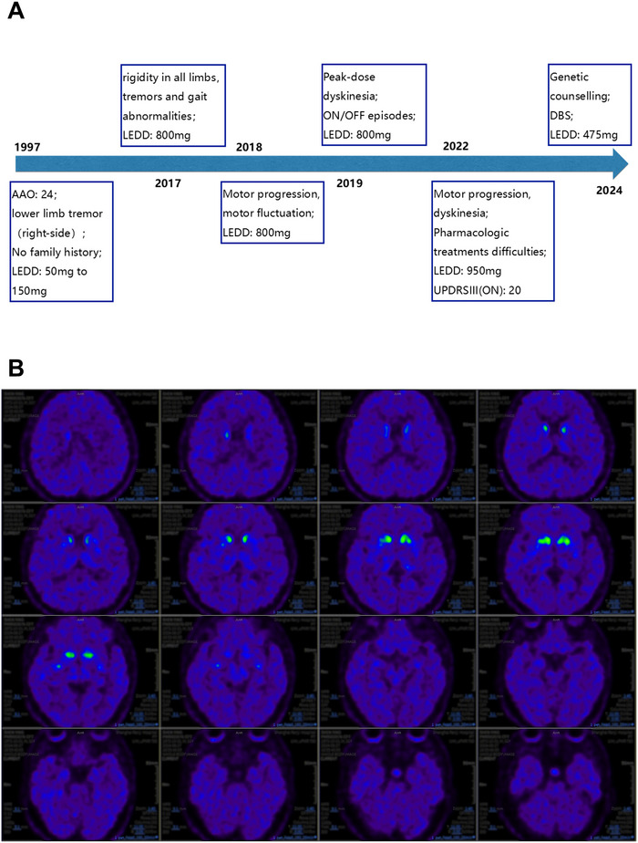
In August 2024, a 51‐year‐old male with a 27‐year history of tremors in his right lower limb presented to the movement disorders clinic at Renji Hospital (Figure 1A). He was born to healthy, normally developing, unrelated parents with a negative family history. Here, we went over his medical history, presented the patient's traits, and identified the associated pathogenesis using genetic testing, biochemical, imaging, and a cell model.
At the age of 24, he acquired resting and postural tremors in his right lower leg, accompanied by a forward‐leaning stride. With 50 mg of levodopa daily, symptoms improved. Over the next decade, his levodopa dose steadily climbed to 150 mg daily (levodopa equivalent daily dose, LEDD, 150 mg) (Jost et al. 2023), and his symptoms remained constant. In 2016, he experienced all‐limb rigidity, obvious tremors, and substantial gait problems. He then upped LEDD to 800 mg on his own, which relieved his symptoms. However, due to emotional turmoil caused by his father's death and a divorce, his symptoms changed. In 2018, he had increased limb rigidity, decreased medicine efficacy, and odd movements during dreams. Dyskinesias developed gradually, lasting about 4–5 h per day during his ON period. When instructed to reduce LEDD to 600 mg, he had uncontrollable tremor and rigidity; therefore, he returned to the previous dose of 800 mg. His dyskinesia worsened dramatically in 2022, and the medication's efficacy fell even more. He tried taking 250 mg of Sinemet at night and taking Pramipexole and Amantadine sporadically, but his symptoms did not get better. The patient has maintained levodopa and Sinemet at an LEDD of 950 mg for the past 2 years since he was unable to tolerate lowering the dosage.

Disease Progression and Treatment of an EOPD Patient. (A) Disease progression: Before 2022, the patient took levodopa 200 mg four times daily; from 2022, 250 mg Sinemet per night was added. AAO: age at onset; LEDD: levodopa equivalent daily dose; DBS: deep brain stimulation, a surgical treatment method; UPDRS III: Unified Parkinson's Disease Rating Scale Part III, used to assess motor function. (B) PET scan: Reduced uptake ofC‐CFT was observed in the bilateral caudate nuclei and putamina. 11
Results
When he first came to us for his ON episode, he had somewhat increased muscle tone in his neck, left upper limb, and both lower limbs, as well as normal muscle strength and coordination. The flexibility of the right lower limb was reduced, as were resting tremors and postural tremors in both upper limbs. Dyskinesia was noticeable, and he walked with a downward tilt of his right shoulder (Video S1). His UPDRS scores (Goetz et al. 2008) of I–IV were 2, 5, 20, and 12, and his Montreal Cognitive Assessment (MoCA) was 21/30, showing modest cognitive loss, particularly in abstraction and delayed recall (Langa and Levine 2014). His other neuropsychological testing was normal and he responded with a 42.2% reduction in UPDRS III scores on the levodopa challenge test.
In terms of neuroimaging, the brain magnetic resonance imaging (MRI) scan returned normal results. A positron emission tomography (PET/MRI) scan, however, revealed increased FDG metabolism in the bilateral thalami and pons, while decreased metabolism was seen in the bilateral medial frontal lobes, bilateral inferior parietal lobes, right inferior frontal lobe, and bilateral lateral temporal lobes. The left dorsal frontal lobes were the most hypometabolic area. Reduced uptake of 11C‐CFT was observed in the bilateral caudate nuclei and putamina (Figure 1B), while the lowest standard uptake value ratio was found in the right posterior putamen (Table S1).
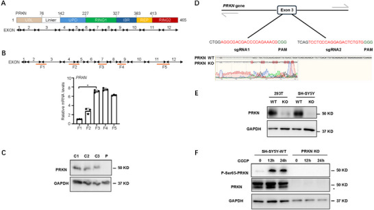
Furthermore, whole‐exome sequencing identified a homozygous exon 3 deletion and a heterozygous exon 4 deletion in PRKN, as well as a heterozygous PINK1 variant NM_032409.3:c.586C>T (p.P196S) in exon 2. To confirm gene expression, we created 5 distinct primer pairs between 12 PRKN exons (methods described in Supporting Information S1). The fragments 1 (F1) and 2 (F2) were reduced in the patient's peripheral blood mononuclear cells (PBMC), which was consistent with the genomic sequencing results (Figure 2B). The PRKN protein was undetectable in the patient's sample (P) compared to the three healthy control samples (C1–C3) (Figure 2C). Meanwhile, we knocked out PRKN using CRISPR‐Cas9 system by targeting exon3 area in 293T and SH‐SY5Y cells (Figure 2D,E) and created an in vitro mitochondrial stress model utilizing CCCP (carbonyl cyanide 3‐chlorophenylhydrazone) therapy at different times in SH‐SY5Y cell (Cook et al. 2024; Geisler et al. 2010). PRKN is downstream of kinase PINK1, so the phosphate of PRKN's serine 65 site increased in SH‐SY5Y WT cells, whereas PRKN and phosphorylated PRKN were undetectable in PRKN knock‐out SH‐SY5Y cells. The in vitro model indicates that PRKN deletion blocks the activation of the PINK1‐PRKN pathway, implying that our patient's mitophagy pathway has been disrupted as a result of PRKN deletion.
Therefore, the diagnosis is EOPD with PRKN/PINK1 mutations. Subthalamic nucleus deep brain stimulation (STN DBS) surgery was proposed and carried out, resulting in significant symptom relief and a 50% reduction in dopaminergic medication (LEDD 950–475 mg) (Video S2).

gene and protein expression. (A) Schematic diagram of PRKN protein functional domains. Human PRKN protein includes 465 amino acids (upper). Correspondingly,gene transcript has 12 exons (lower). (B) The regions of five pairs of primers are shown in red line under the 12 exons ofgene transcript diagram, annotated with F1–F5 (upper). The relative mRNA expression level of five fragments in patient's PBMC is shown in the bar chart (lower). Data represent the mean ± SEM. (C) PRKN protein level in three control samples (C1–C3) and patient (P). PBMCs were isolated from three normal individuals and patient. (D) Theknockout strategy is shown in 293T and SH‐SY5Y cell lines (upper); Sanger sequencing was used to detect the knockout fragment ofin SH‐SY5Y cell line (lower). (E) PRKN protein knockout efficiency in 293T and SH‐SY5Y cell lines using western blot. (F) The PRKN phosphorylation of ser65 site was detected by western blot after 20 uM CCCP treatment under indicated conditions. The non‐specific band of PRKN is marked with a black asterisk. PRKN PRKN PRKN PRKN PRKN
Discussion
The case presented a PRKN/PINK1‐related EOPD with a lower AAO, slower development, early dyskinesia, and a favorable response to levodopa and STN DBS. Otherwise, it is puzzling that PRKN or PINK1‐PD caused different pathologies of dopaminergic neuronal loss and Lewy bodies, despite the fact that PRKN and PINK1 both sent disabled mitochondria to the lysosome for breakdown during mitophagy (Han et al. 2023).
Fewer studies have examined the relationship between the variations and structural/functional deficits to date, particularly when a patient has numerous mutations. Our case not only revealed unusual double mutations of PRKN/PINK1 by WES, but also confirmed gene and protein expression (Figure 2). Only three examples in the Polish literature had previously been documented to have simultaneous mutations in the PRKN/PINK1 genes; however, the mutation patterns differ from those seen in our cases (Milanowski et al. 2021). Furthermore, two other research works focusing on the North American and Chinese cohorts did not find similar multigene alterations (Cook et al. 2024; Sun et al. 2023). The patterns of multigene changes in PRKN/PINK1 and their relationship with mitophagy require more investigation. Our findings indicated that the exon 3 deletion of PRKN in the patient, which was the main structural variant (located in the RING0 protein domain and the ubiquitin‐like protein domain), likely prevented its activation (Menon et al. 2024). The patient's motor symptoms are most likely caused by damaged mitophagy via the PINK1‐PRKN pathway (Geisler et al. 2010; Han et al. 2023). Our patient's unusual cognitive impairment could be attributed to FDG hypometabolism in the bilateral medial frontal and inferior parietal lobes, as seen in other cases with Lewy body illness (Ferreira et al. 2025). Our findings indicate that PRKN/PINK1‐PD is a hybrid of defective mitochondria and alpha‐synuclein aggregation, as previously shown in cell and animal models (Hou et al. 2023).
Conclusion
Genetic data are essential for creating gene‐specific trials because PRKN/PINK1‐PD has a better prognosis and an earlier AAO than sporadic PD. We propose providing a PD gene panel with PRKN/PINK1 to EOPD patients, particularly those with AAO < 40 years of Asian heritage. More research is needed to confirm the phenotype–genotype links underlying the PINK1‐PRKN pathway and create gene‐specific treatments.
Author Contributions
Zhongjiao Lu: conceptualization, funding acquisition, investigation, resources, writing – original draft, writing – review and editing. Ye Sun: investigation, resources, data curation, formal analysis, funding acquisition. Liemei Guo: conceptualization, investigation, resources. Mei Xin: investigation, resources. Yunlan Du: investigation, resources, data curation, formal analysis. Ruolian Dai: investigation, resources, data curation, formal analysis. Gang Chen: resources, investigation. Hualong Wang: methodology, writing – review and editing. Gang Wang: conceptualization, methodology, funding acquisition, writing – review and editing.
Disclosure
The authors have nothing to report.
Ethics Statement
The clinical study protocol and the informed consent form were reviewed and approved by the independent ethics committees of the participating sites. All participants gave full written consent, and the patient agreed to genetic analysis for diagnostic and scientific purposes and for online video publication. We confirm that we have read the Journal's position on issues involved in ethical publication and affirm that this work is consistent with those guidelines.
Conflicts of Interest
The authors declare no conflicts of interest.
Peer Review
The peer review history for this article is available at https://publons.com/publon/10.1002/brb3.70822↗.