
What this is
- This research explores a novel genome editing tool, the Ago-F system, for Mycobacterium smegmatis.
- Mycobacterium smegmatis serves as a model organism for studying tuberculosis due to its nonpathogenic nature.
- The study demonstrates high editing efficiency and reduced time requirements for gene knockout, addressing limitations in current genetic tools.
Essence
- The Ago-F system achieves 80% knockout efficiency in Mycobacterium smegmatis, significantly enhancing genome editing capabilities in this model organism.
Key takeaways
- The Ago-F system enables efficient genome editing in Mycobacterium smegmatis, achieving an editing efficiency of 80%. This level of efficiency is critical for advancing functional genomics research.
- The time required for genome editing was reduced to 8 days with the Ago-F system, streamlining the process compared to traditional methods.
Caveats
- The study relies on Mycobacterium smegmatis, which may not fully represent the complexities of pathogenic mycobacteria like Mycobacterium tuberculosis.
- While the editing efficiency is promising, the long-term effects and stability of the edits in subsequent generations were not addressed.
Simplified
INTRODUCTION
Tuberculosis (TB), caused by Mycobacterium tuberculosis, is a highly lethal disease that is transmitted primarily through the respiratory tract. According to the "Global Tuberculosis Report 2024," released in 2024, the global death toll from tuberculosis reached 1.25 million, replacing COVID-19 as the world's leading cause of death from a single infectious agent. Research on the mechanism of pathogenicity in M. tuberculosis is currently a key focus in tuberculosis prevention and control efforts (1). However, current limitations in mycobacterial genetic tools, particularly low editing efficiency and technical constraints, pose significant barriers to studying TB pathogenesis and developing effective interventions. To address these methodological challenges, the nonpathogenic model organism M. smegmatis was selected for this study due to its unique experimental advantages. Its fast growth cycle, genetic accessibility, and safe handling characteristics make M. smegmatis exceptionally suitable for developing and optimizing improved genetic manipulation systems (2). Establishing efficient and universal genetic manipulation platforms using M. smegmatis is expected to substantially accelerate investigations into M. tuberculosis pathogenesis-related gene functions and provide critical technical support for the development of novel anti-TB strategies, complementing direct studies in M. tuberculosis.
Traditional genome editing methods for M. smegmatis rely on allele exchange, homologous recombination, and nonhomologous end joining (NHEJ), but the knockout efficiency is very low (10−5‒10−6) (3). With the development of CRISPR technology, CRISPR/dCas9 has been shown to reduce gene expression in M. smegmatis (4). The CRISPR-Cas12a-assisted homologous recombination system was subsequently used for genome editing in M. smegmatis, and the efficiency of introducing site-specific point mutations reached 80%, whereas the deletion/insertion efficiency of double-stranded DNA recombination was 37%–75% (5). Recent studies have shown that the CRISPR-FnCpf1-assisted NHEJ system can repair double-strand breaks (DSBs) in M. smegmatis without requiring recombinant proteins or homologous DNA templates, enabling single-gene knockout with a total time requirement of 7 days (6). However, although this CRISPR-mediated knockout system provides a tool for rapid and efficient mycobacterial genome modification, challenges associated with its use exist, such as the complexity of the construction of plasmids harboring sgRNA, varying efficiencies of different sgRNAs, off-target effects, and the large size of cas protein-containing plasmids, leading to difficulties in vector construction and transformation. These challenges limit the application of this system in actinomycetes, in which plasmid transformation is difficult. The development of a simple and efficient tool for rapid genome editing could accelerate functional genomic investigations of M. smegmatis and be beneficial for exploring the mechanism of M. tuberculosis pathogenesis.
The Argonaute (Ago) protein is a programmable nucleic acid enzyme found in both eukaryotes and prokaryotes. Studies have shown that while prokaryotic Ago protein (pAgo) and eukaryotic Ago protein (eAgo) share high structural homology, eAgo proteins can cleave only RNA targets, whereas many pAgo proteins can cleave DNA targets (7). In prokaryotes, Agos are divided into long pAgos and short pAgos, among which long pAgos consist of four domains, namely, the N-terminal, PIWI–Argonaute–Zwille (PAZ), middle (MID), and PIWI domains, as well as the L1 and L2 linkers, whereas short pAgos consist of only the MID and PIWI domains (7–9). Many pAgos, such as TtAgo (10), MjAgo (11), and PfAgo (12), can function only at temperatures above 65°C with gDNA as the guide. However, NgAgo was found to function at 30°C or 37°C, and this temperature is suitable for the growth of most organisms (8, 13). In 2016, Gao et al. (13) discovered that NgAgo functions as a DNA-guided endonuclease and is applicable for genome editing in human cells. However, subsequent studies by other research teams were unable to confirm that NgAgo possesses gene-editing capabilities. In the same year, Qi et al. reported that gDNA/NgAgo can bind to target genes to block transcription in zebrafish, a eukaryotic organism, but no gene-editing activity of NgAgo was observed (14). In 2019, Fu et al. confirmed that NgAgo physically interacts with RecA to mediate homologous recombination in bacteria (8). In 2021, Lee et al. confirmed that NgAgo has guided DNA nicking activity and further demonstrated that NgAgo performs targeting and gene editing in E. coli (15). In 2022, Xing et al. reported that NgAgo had DNA cleavage activity similar to that of Cas9 (16). Although NgAgo cannot knock out genes in eukaryotic organisms, it can function in prokaryotes, such as Pasteurella multocida and Escherichia coli, through RecA-mediated recombination (8). The prokaryotic-specific activity of NgAgo arises from its dependence on RecA, compatibility with bacterial DNA repair pathways, and evolutionary optimization for prokaryotic physiology. In contrast, eukaryotic barriers—including incompatible repair mechanisms and host immune responses—preclude editing. In addition, researchers have analyzed the truncated domains of the NgAgo protein and reported that the last 717 bp of the PIWI domain, namely, NgAgo-F, is the primary domain that mediates bacterial genome editing (8). NgAgo-F can interact with the intracellular homologous recombination enzyme RecA, increasing the RecA-mediated homologous DNA strand exchange capability and thereby increasing recombination efficiency, and the knockout efficiency can reach 80%–100% (8). In this study, the NgAgo-F system was developed for genome editing in M. smegmatis. The constitutive promoter of a heat shock protein, Hsp60 (6), was used to drive NgAgo-F expression in pKC1139 (17). GlnR, a global transcriptional regulator of nitrogen metabolism, and LtmA, a receptor for c-di-GMP, were chosen as the targets, and the upstream and downstream homology arm sequences were then inserted into pKC1139. After transformation into M. smegmatis, qRT-PCR and sequencing were performed to identify whether the glnR or ltmA genes were successfully knocked out. To further reduce the plasmid size, we compared the effects of homology arm length on the knockout efficiency of the glnR gene in M. smegmatis. The 200 bp homologous arm sequences were sufficient to successfully knock out the glnR gene. In summary, the NgAgo-F-based system provides a quick, efficient, and easy-to-use gene editing tool for use in M. smegmatis, with significant potential for application in tuberculosis prevention and treatment research.
RESULTS
Plasmid construction
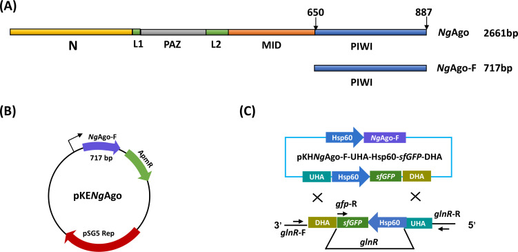
NgAgo-F is the PIWI domain of NgAgo, which is approximately 717 bp in length and mediates bacterial genome editing (Fig. 1A). We codon-optimized the sequence of the NgAgo-F gene to make NgAgo-F much easier to express and function in M. smegmatis. Using homologous recombination, we successfully inserted Flag-NgAgo-F driven by the Hsp60 promoter into the shuttle plasmid pKC1139, which contains a temperature-sensitive pSG5 replication system and a cassette encoding the aac(3)-Ivan gene for apramycin resistance that can be subsequently lost through temperature treatment (18). Through this method, the pKC1139-Hsp60-Flag-NgAgo-F (pKHNgAgo-F) plasmid was constructed (Fig. 1B). Then, upstream and downstream sequences, with superfolder green fluorescent protein (sfGFP) between them, were introduced into the pKHNgAgo-F plasmid. A schematic diagram of the constructed knockout plasmid and the knockout principle is shown in Fig. 1C.

Construction of knockout-related plasmids. () Domain organization ofAgo andAgo-F. () Composition of theAgo-F-mediated plasmids. A FlagAgo-F gene with an60 promoter was introduced into the plasmid pKC1139 to generate the plasmid pKHAgo-F. () Scheme ofgene knockout invia theAgo-F system. A B C Ng Ng Ng -Ng Hsp Ng glnR M. smegmatis Ng
Solubility verification analysis ofAgo-F Ng
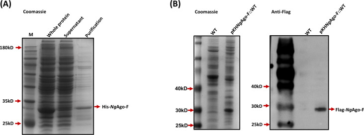
The solubility of the NgAgo-F protein is critical to its function. To verify the solubility of NgAgo-F, we first constructed the pET28a-His-NgAgo-F plasmid via the homologous recombination method. The plasmid was subsequently transformed into BL21(DE3) cells. His-NgAgo-F, a 31.2 kDa fusion protein, was successfully expressed and purified (Fig. 2A), demonstrating that NgAgo-F is soluble in E. coli. To further confirm whether the NgAgo-F protein is soluble in M. smegmatis, we transferred pKHNgAgo-F into the wild-type (WT) strain and obtained the strain pKHNgAgo-F::WT. Western blotting was performed using an anti-Flag antibody. The NgAgo-F protein was detected in the supernatant, confirming that the NgAgo-F protein is also soluble in M. smegmatis (Fig. 2B). These results indicate that NgAgo-F can achieve genome editing in M. smegmatis.

Ago-F is soluble inand. () His-Ago-F protein expression and purification in BL21(DE3) cells. The His-Ago-F protein was separated by 15% SDS‒PAGE and stained with Coomassie Brilliant Blue R-250. () The Flag-Ago-F protein was expressed inand identified via western blotting with an anti-Flag antibody. Left: Coomassie blue staining of supernatant protein from WT and pKHAgo-F::WT. Right: Western blotting measurement ofAgo-F in the supernatants of WT and pKHAgo-F::WT. Ng E. coli M. smegmatis Ng Ng Ng M. smegmatis Ng Ng Ng A B
Deletion ofin glnR M. smegmatis
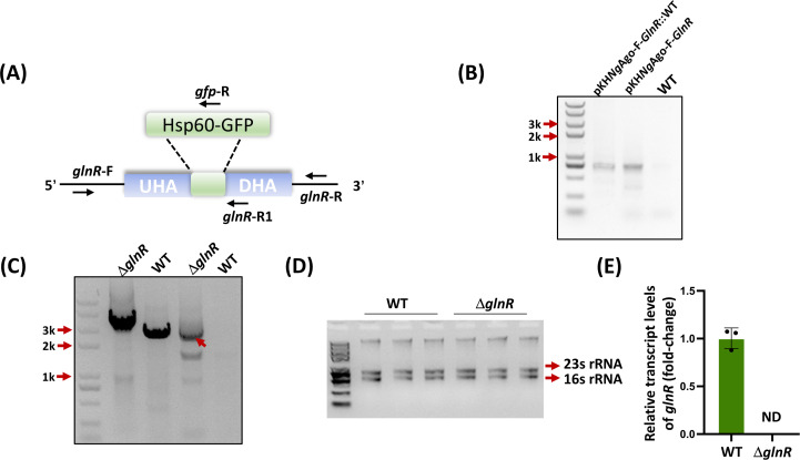
To determine whether the NgAgo-F tool can achieve genome deletion in M. smegmatis, we first constructed a plasmid for glnR knockout. To ensure high cleavage efficiency, we recombined 800 bp upstream and 800 bp downstream homology arms into the pKHNgAgo-F plasmid and obtained the plasmid pKHNgAgo-F-GlnR. After the plasmid was transformed into M. smegmatis through electroporation for 3‒4 days, individual colonies were transferred to liquid LB media for further cultivation, and PCR was performed to confirm that the plasmids were successfully transferred into M. smegmatis (Fig. 3B). After three passages, PCR was performed, and the products were sequenced to further confirm that the glnR gene was indeed knocked out in strains with successfully transferred plasmids (Fig. 3A and C). Additionally, we extracted RNA from the WT and ΔglnR strains and conducted qRT‒PCR analysis. Our results revealed that in the ΔglnR mutant, the expression of glnR was absent, which indicated that glnR was knocked out successfully and that the NgAgo-F system can be used for genome editing in M. smegmatis (Fig. 3D and E).

deletion in. ()in genomic DNA was replaced with60-. () Plasmid pKHAgo-F-was transformed into WT through PCR identification. Plasmid pKHAgo-F-and WT strain represent positive and negative controls, respectively. ()knockout identification of the strains via PCR amplification using the primers-F/R (left two lines) and-F/-R (right two lines), with Δand WT as the templates. () Agarose gel electrophoresis of total RNA (with genomic DNA) extracted from WT and Δ, and the resulting RNAs were not degraded. () Relative expression ofin the WT and Δstrains. ND: not detected. The error bars indicate the SDs of three independent biological experiments, and the mean values were calculated on the basis of these replicates. glnR M. smegmatis glnR Hsp GFP Ng GlnR Ng GlnR glnR glnR glnR gfp glnR glnR glnR glnR A B C D E
Deletion ofin ltmA M. smegmatis
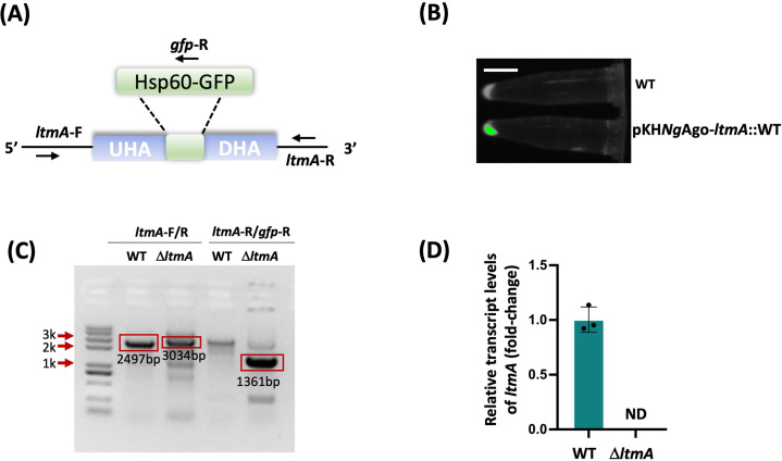
To test whether NgAgo-F-based gene knockout in M. smegmatis could be applied to genes other than glnR, we applied this approach to manipulate another gene, ltmA. We recombined the 800 bp upstream and downstream homology arms of the ltmA gene and sfGFP fragment into the pKHNgAgo-F vector, obtaining the plasmid pKHNgAgo-F-ltmA. The plasmid was then transferred into M. smegmatis, and single colonies were selected for cultivation. Through fluorescence signals, we identified the strains that were successfully transformed with the plasmid pKHNgAgo-F-ltmA (Fig. 4B). After three passages, we confirmed the deletion of ltmA via PCR amplification with the primers ltmA-F/R and ltmA-R/gfp-R and obtained the ΔltmA mutant (Fig. 4A and C). To further substantiate the knockout results, RNA was extracted, and qRT-PCR analysis revealed that the expression of ltmA was eliminated in the ΔltmA mutant (Fig. 4D). These results further support that the NgAgo-F system can be used for genome editing in M. smegmatis.

deletion in()in the genomic DNA was replaced with60-. () The fluorescence signal suggested that the plasmid withwas successfully transformed intoThe signal was captured via a ChemiScope 6100 chemiluminescence imaging system. Scale bars, 1 cm. () PCR-mediated identification of the recombinant strains via the-F/R and-F/-R primers. The 2,497 bp band is the length amplified via the primers-F/R with the WT as the template. The 3,034 bp band is the length amplified via the primers-F/R with the Δmutant template. The 1,361 bp band is the length amplified via the primers-R/-R with the Δmutant. () Relative expression ofin the WT and Δstrains. ND: not detected. The error bars indicate the SDs of three independent biological experiments, and the mean values were calculated on the basis of these replicates. ltmA M. smegmatis ltmA Hsp GFP GFP M. smegmatis. ltmA ltmA gfp ltmA ltmA ltmA ltmA gfp ltmA ltmA ltmA . A B C D
Influence of homology arm length onAgo-F-assisted gene editing efficiency Ng
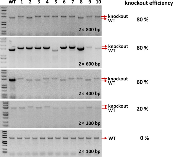
Plasmid transformation is a necessary step for gene editing. The larger the plasmid is, the lower the transformation efficiency. For some difficult-to-transform strains, a small plasmid is considered essential for successful transformation. To further screen for small plasmids for NgAgo-F-mediated gene editing in M. smegmatis, we explored the impact of homology arm length on NgAgo-F cleavage activity in M. smegmatis, using glnR as the target. We constructed glnR gene knockout plasmids with homology arm lengths of 2 × 100 bp, 2 × 200 bp, 2 × 400 bp, 2 × 600 bp, and 2 × 800 bp, and separately transformed them into M. smegmatis to calculate the gene knockout efficiency (Fig. 5). The results indicated that the construction with a 2 × 100 bp homology arm length could not completely knock out the gene; a 20% knockout efficiency was achieved with a 2 × 200 bp homology arm length; a 60% knockout efficiency was achieved with a 2 × 400 bp homology arm length; an 80% knockout efficiency was achieved with a 2 × 600 bp homology arm length; and an 80% knockout efficiency was achieved with a 2 × 800 bp homology arm length (Fig. 5). Through comparison, we found a 600 bp homology arm length is sufficient to achieve efficient gene knockout, and that the minimum length of the knockout plasmid could be reduced to 200 bp. Reducing the length of the plasmid increased the likelihood of successful plasmid transformation into mycobacteria, leading to successful knockout of the target gene.

Impact of arm length on theAgo-F-assisted gene editing efficiencyComparison of the knockout efficiency of themutant inon the basis ofAgo-F with different lengths of the homology arm. Ten clones were used to calculate the knockout efficiency with the primers-F/R1. Ng glnR M. smegmatis Ng glnR .
DISCUSSION
To our knowledge, the NgAgo-F system developed for M. smegmatis transformation in this study is easy to use and has minimal restrictions for the recognition of sequences without a PAM site. The adaptability of NgAgo-F to mycobacterial genomic constraints highlights its potential to accelerate functional studies of essential metabolic pathways. For example, glnR knockout strains could elucidate the role of nitrogen assimilation in mycobacterial survival, whereas ltmA deletion may reveal lipid homeostasis mechanisms critical for pathogenicity in related species. The enhanced efficiency and reduced timeline address a critical bottleneck in mycobacterial genetics, enabling rapid validation of gene‒phenotype relationships. Moreover, the plasmid simplicity and GC-rich targeting flexibility of the NgAgo platform suggest broader applicability to other Actinobacteria, which are notoriously recalcitrant to genetic manipulation. By streamlining genome editing, this approach may expedite the identification of therapeutic targets or resistance mechanisms in pathogenic mycobacteria, ultimately supporting the development of novel antimicrobial strategies.
MATERIALS AND METHODS
Strains, plasmids, and culture conditions
E. coli DH5α served as the host for plasmid construction and was used to assess NgAgo-F-assisted gene editing efficiency. E. coli BL21(DE3) was used for protein expression and purification. Additionally, M. smegmatis MC2 155 served as the host for evaluating NgAgo-F-assisted gene editing. Unless otherwise stated, E. coli DH5α, BL21(DE3), and M. smegmatis were grown in LB media at 37°C and 220 rpm, with appropriate antibiotics added to the broth as needed.
Plasmid construction
The NgAgo-F gene was synthesized by Sangon Biotech Co., Ltd. (Shanghai, China), with codon optimization. To construct the pKHNgAgo-F plasmid, the restriction enzymes EcoR I and EcoR V were used to obtain the pKC1139 vector. The Flag-NgAgo-F fragment and Hsp60 promoter were amplified via the primers Flag-NgAgo-F/R and Hsp60-F/F1/R. These two fragments have 25 bp overlapping sequences, and the two fragments were subsequently fused through PCR amplification. The fused fragment was connected to the pKC1139 vector via the pEASY-Basic Seamless Cloning and Assembly Kit (Transgene, Beijing, China). The upstream and downstream homology arms of the glnR gene and ltmA gene were amplified using M. smegmatis genomic DNA as the template. These homology arms were separately cloned and inserted into the pKHNgAgo-F vector, generating the plasmids pKHNgAgo-F-glnR and pKHNgAgo-F-ltmA. Finally, pKHNgAgo-F-glnR and pKHNgAgo-F-ltmA were transformed into M. smegmatis for further applications.
Preparation of competentcells and electroporation methods M. smegmatis
M. smegmatis was cultured in LB liquid media supplemented with 0.05% Tween 80 at 37°C for 2 days. Subsequently, 500 µL of the culture was added to 100 mL of LB liquid media containing 0.05% Tween 80 in a 500-mL shake flask and incubated at 37°C with shaking at 220 rpm for approximately 36 h. When the OD600 reached 0.6, the cells were immediately placed on ice and incubated for 30 min. The cells were harvested and collected by centrifugation at 6,000×g at 4°C for 10 min. After the supernatants were removed, the cells were washed once with cold water and washed twice with cold 10% glycerol. Finally, the cells were resuspended in 500 µL of 10% glycerol, and 200 µL aliquots were divided into 1.5 mL sterile centrifuge tubes.
For electroporation, 20 µL of plasmid DNA was added to 200 µL of competent cells, and the mixture was transferred into precooled 2-mm electroporation cuvettes. Electroporation was performed at 3 kV for 4 ms once or twice via a MicroPulser (Bio-Rad, 411BR7769). Immediately, 900 µL of LB medium was added to the competent cells, and the mixture was incubated with shaking at 30°C and 220 rpm for approximately 3 h. Following recovery, the cells were centrifuged, and the cells in the final 200 µL were resuspended and plated on LB agar plates containing 50 µg/µL apramycin. The plates were incubated for 3‒4 days. To ensure transformation efficiency, competent cells should be prepared and used immediately.
Protein expression and purification
The primers 28a-NgAgo-F-F/R were used to amplify the NgAgo-F fragment, and the primers pET28a-F/R were used to amplify the pET28a vector fragment. The two fragments had 25 bp overlapping sequences, and the overlapping sequences were designed with the primers pET28a-F/R. In the pEASY-Basic Seamless Cloning and Assembly Kit, a basic mixture was used. The assembly system contained 6 µL of Basic Mix, x µL of NgAgo-F fragment, and (6−x) µL of pET28a vector, and the mixture was incubated at 50°C in a water bath for 30 min. The molar ratio of the NgAgo-F fragment to the pET28a vector was 3:1‒5:1. The mixture was subsequently transformed into competent cells. T7 and T7 Ter primers were used to identify the correct clones. The recombinant plasmid pET28a-NgAgo-F was transformed into E. coli BL21(DE3) competent cells for protein expression. The transformed cells were initially cultured in LB liquid media at 37°C overnight. Then, 5 mL was added to 100 mL of LB liquid media in a 500 mL shake flask and cultured at 37°C with shaking at 220 rpm for approximately 2‒3 h. When the OD600 reached 0.6, His-NgAgo-F was induced by adding 0.5 mM IPTG, followed by incubation at 18°C for 14‒16 h. The cells were collected by centrifugation at 6,000×g for 10 min and washed once with 1× PBS. After being resuspended in 35 mL of 1× PBS, the cells were lysed via sonication, and the supernatants were collected via centrifugation at 9,000×g for 20 min at 4°C. Protein purification was performed according to previously established protocols (19). The purified protein was analyzed by 15% SDS‒PAGE (Epizyme Biotech, Shanghai, China).
Western blotting
pKHNgAgo-F::M. smegmatis and M. smegmatis were cultured in LB liquid media supplemented with 0.05% Tween 80 at 30°C for 2 days. The cells were collected by centrifugation at 6,000×g for 20 min and washed once with 1× PBS. The cell pellet was resuspended in 300 µL of 1× PBS and lysed by sonication, and the supernatants were collected by centrifugation at 9,000×g for 20 min at 4°C. The supernatants of pKHNgAgo-F::M. smegmatis and M. smegmatis were mixed with 6× protein loading buffer and run on SDS‒PAGE gels. Next, the proteins on the SDS‒PAGE gels were transferred to a PVDF membrane (Merck, Millipore) at 300 V and 380 mA for 90 min. After being washed twice with TBST buffer, which consisted of 20 mM Tris-HCl, pH 7.6, 150 mM NaCl, and 0.1% (v/v) Tween 20, the PVDF membrane was incubated with 0.5% BSA blocking buffer at 4°C overnight. Two microliters of anti-Flag antibody (Transgene, Beijing, China) was added to the BSA blocking buffer and incubated at 25°C for 1.5 h. Then, the PVDF membrane was washed three times with TBST buffer, and 2 µL ProteinFind Goat Anti-Mouse IgG (H + L) (Transgene, Beijing, China) was added to 10 mL of TBST buffer. After incubation for approximately 1 h, the PVDF membrane was washed three times with TBST, and an enhanced chemiluminescence (ECL) system (CTB, USA) was used to detect the signal.
qRT‒PCR
WT, ΔglnR, and ΔltmA were cultured in LB media supplemented with 0.05% Tween 80 at 37°C for 2 days. Total RNA was extracted from WT, ΔglnR, and ΔltmA strains with an RNAprep Pure Cell/Bacteria Kit (Tiangen Biotech, Beijing, China) according to the manufacturer's instructions. cDNA was subsequently synthesized via TransScript Uni All-in-One First-Strand cDNA Synthesis SuperMix (Transgene, Beijing, China) for qRT‒PCR. A 96-well reaction was performed with a Bio-Rad CFX96 real-time PCR detection system (Bio-Rad, Hercules, CA, USA) using TB Green Premix Ex Taq (TaKaRa Biotechnology, Beijing, China). All PCR mixtures were prepared in a volume of 20 µL in triplicate and contained 10 µL of SYBR Green PCR master mix, 0.4 µM forward primer, and 0.4 µM reverse primer. The PCR conditions were as follows: 95°C for 5 min, followed by 40 cycles of 95°C for 30 s, 55°C for 20 s, 72°C for 15 s, and 72°C for 10 min. The expression levels of glnR and ltmA were normalized to that of the housekeeping gene sigA (MSMEG_2758).